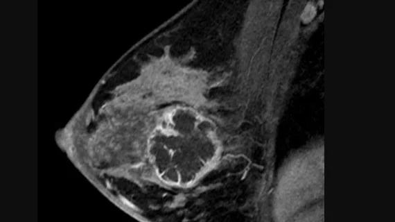
Researchers identify MRI findings linked with invasive breast cancer
Focal edema observed on breast MRI exams could signal a more invasive cancer diagnosis for women who display such findings.
According to a new paper published in Insights into Imaging, focal edema on T2-weighted images (T2WI) was recently found to positively correlate with several factors indicative of a tumor’s invasiveness. The authors of the new paper suggested that this understanding could serve as a noninvasive tool in creating more personalized treatment options for patients facing a cancer diagnosis that is invasive in nature.
“Various features extracted from breast MRI have the potential to serve as noninvasive biomarkers that could predict the biologic behavior of the cancer,” corresponding author Yuzhen Zhang, with the Department of Radiology at Xinhua Hospital in China, and co-authors noted. “In current research, the dynamic contrast-enhanced magnetic resonance imaging and diffusion weighted imaging techniques are hotspots. By contrast, the research on the T2 fat suppression sequence has been less extensive.”
The authors pointed to several potential benefits of T2WI sequences, including increased specificity in differentiating between benign and malignant lesions and superior visualization of breast edema. While edema of the breast has been linked with tumor aggressiveness in the past, its exact prognostic value is not yet completely understood.
The team sought to better define the relationship between breast edema on MRI and cancer invasiveness. To do this, they retrospectively analyzed the cases of 201 patients diagnosed with invasive breast cancer, comparing their biopsy-confirmed diagnoses with image features and breast edema scores (BES) derived from their breast MRI exams.
On the fat-suppressed T2WI sequences, focal edema was observed in 102 out of 205 lesions. This feature was found to be positively correlated with several characteristics of biologically aggressive cancer—larger tumors (2 cm or more), patients with higher histologic grade, negative ER status, negative PR status, higher Ki-67 index and positive HER2 status. Additionally, higher BES, higher rates of axillary lymph node metastases and non-luminal breast cancer were also associated with the presence of focal edema.
Combined, the team suggested that these features “could predict worse prognosis and probably contribute to disease recurrence.”
“As a potential imaging biomarker for the comprehensive risk classification and prognostication for patients with breast cancer, BES may be valuable for defining the appropriate therapeutic targets for patients who need a more intensive adjuvant management,” the group explained.
Despite some acknowledged study limitations, the authors maintained that these preoperative MRI findings could be used as practical tool to reliably guide treatment decisions by better informing both clinicians and patients.
The study abstract is available here.

