Breast Imaging

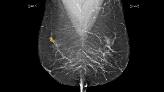
Breast imaging includes imaging modalities used for breast cancer screenings and planning therapy once cancer is detected. Mammography is the primary modality used. Mammogram technology is moving from 2D full-field digital mammography (FFDM) to breast tomosynthesis, or 3D mammography, which helps reduce false positive exams by allowing radiologists to look through the layers of tissue. Overlapping areas of dense breast tissue on 2D mammograms appear similar to cancers and 3D tomo helps determine if suspect areas are cancer or not. About 50% of women have dense breast tissue, which appears white on mammograms, the same as cancers, making diagnosis difficult. Radiologists use the Breast Imaging Reporting and Data System (BI-RADS) scoring system to define the density of breast tissue. Many states now require patients to be notified if they have dense breasts so they understand their mammograms might be suboptimal and they should use supplemental imaging that can see through the dense areas. This includes tomosythesis, breast ultrasound, automated breast ultrasound (ABUS), breast MRI, contrast enhanced mammography and nuclear imaging, including positron emission mammography (PEM).

breast radiologist breast cancer mammography

Radiation treatment for breast cancer linked to high risk of developing subsequent skin cancer

The danger of developing the disease on or around the breast after radiation is 57% higher than the general population.

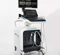
Seno Medical Imagio breast imaging system.

AMA adds code to allow for reimbursement of opto-acoustic imaging modality

The new imaging modality was pioneered by Seno Medical for diagnosing breast cancer.

breast cancer women's health ribbon

ESR partners with GE HealthCare to focus on breast cancer treatment

A mobile screening system for breast cancer will be launched at the ECR 2024 conference.

breast cancer mammography screening mammogram scheduling

Arizona bill would eliminate out-of-pocket costs for breast cancer screenings

Susan G. Komen, a breast cancer advocacy group, is urging lawmakers to pass the legislation.

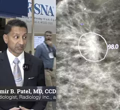
Video of Samir Patel, MD, diagnostic radiologist at Radiology Inc., value management program founder and director and a board member of the Beacon Health System, explaining how AI is being implemented in mammography at RSNA 2023.

Real-world implementation of AI in mammography

Samir Patel, MD, radiologist at Radiology Inc., and a board member of the Beacon Health System, explains how AI is being implemented in clinical practice for breast imaging. 


fingerprint

Your fingerprint sweat may reveal if you have breast cancer

Scientists may have turned their criminal forensics research into an accurate alternative to screening mammography. 

artificial intelligence healthcare industry digest

ChatGPT 'mostly' accurate when answering questions on breast biopsies, with 1 major exception

Researchers from Johns Hopkins University asked the chatbot about common findings from core-needle biopsies.

Thumbnail

Mayo Clinic rings in the new year with two AI-powered cancer care initiatives

Two AI developers have announced new agreements with the academic medical center, both related to improving the delivery of oncology services.

Around the web

The nuclear imaging isotope shortage of molybdenum-99 may be over now that the sidelined reactor is restarting. ASNC's president says PET and new SPECT technologies helped cardiac imaging labs better weather the storm.

CMS has more than doubled the CCTA payment rate from $175 to $357.13. The move, expected to have a significant impact on the utilization of cardiac CT, received immediate praise from imaging specialists.

The newly cleared offering, AutoChamber, was designed with opportunistic screening in mind. It can evaluate many different kinds of CT images, including those originally gathered to screen patients for lung cancer. 

Trimed Popup
Trimed Popup