Breast Imaging

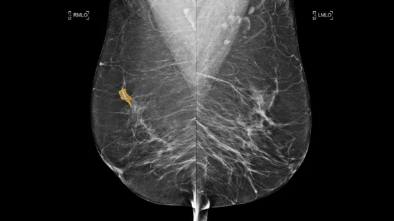
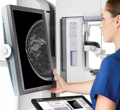
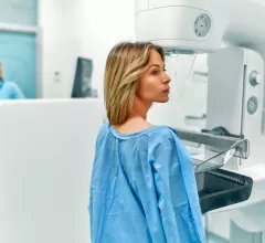
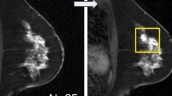
Breast imaging includes imaging modalities used for breast cancer screenings and planning therapy once cancer is detected. Mammography is the primary modality used. Mammogram technology is moving from 2D full-field digital mammography (FFDM) to breast tomosynthesis, or 3D mammography, which helps reduce false positive exams by allowing radiologists to look through the layers of tissue. Overlapping areas of dense breast tissue on 2D mammograms appear similar to cancers and 3D tomo helps determine if suspect areas are cancer or not. About 50% of women have dense breast tissue, which appears white on mammograms, the same as cancers, making diagnosis difficult. Radiologists use the Breast Imaging Reporting and Data System (BI-RADS) scoring system to define the density of breast tissue. Many states now require patients to be notified if they have dense breasts so they understand their mammograms might be suboptimal and they should use supplemental imaging that can see through the dense areas. This includes tomosythesis, breast ultrasound, automated breast ultrasound (ABUS), breast MRI, contrast enhanced mammography and nuclear imaging, including positron emission mammography (PEM).

Common cancer screenings may not lead to longer lives, with 1 key exception

New research out of Norway focused on 6 common cancer screening tests, tracking their impact on more than 2 million patients.

money business cash flow dollar

Dense breast imaging specialists secure $12M in funding

Delphinus Medical Technologies says it will use the investment to expand the commercial adoption of its SoftVue 3D Whole Breast Tomography System.

Thumbnail

New AI model helps radiologists ID breast cancer lesions on ultrasound images

Using the new-look algorithm consistently improved reader accuracy, especially among less experienced radiologists. It also helped limit false-positive results. 

 background parenchyma enhancement breast MRI

MRI finding linked to heightened cancer risk among women with very dense breasts

A new AI model helped researchers identify a relationship between background parenchyma enhancement on breast MRI exams and breast cancer risk.

Thumbnail

Deep learning detects precancerous changes on mammograms

What’s more, the model proved beneficial in a large, diverse cohort that included women with benign breast disease and BRCA mutations.

Thumbnail

Some radiologists' stress lowered during COVID, but it was short lived

Breast radiologists are known to have higher rates of burnout in comparison to many of their peers, but the beginning of the pandemic brought this group something not often encountered within the specialty—downtime. 

mammogram mammography breast cancer

Continued declines in screening mammogram volumes could have 'worrisome implications,' experts warn

The downward trend is most notable in women who have at least one risk factor of severe COVID, new data suggest.

Considerations for supplemental imaging should extend beyond breast density, analysis suggests

Women with increased breast density are targeted significantly more for supplemental imaging than those without dense tissue, but there are other factors that increase the risk of mammography screening failure that should also be considered, according to new data.

Around the web

The nuclear imaging isotope shortage of molybdenum-99 may be over now that the sidelined reactor is restarting. ASNC's president says PET and new SPECT technologies helped cardiac imaging labs better weather the storm.

CMS has more than doubled the CCTA payment rate from $175 to $357.13. The move, expected to have a significant impact on the utilization of cardiac CT, received immediate praise from imaging specialists.

The newly cleared offering, AutoChamber, was designed with opportunistic screening in mind. It can evaluate many different kinds of CT images, including those originally gathered to screen patients for lung cancer. 

Trimed Popup
Trimed Popup