Breast Imaging

Breast imaging includes imaging modalities used for breast cancer screenings and planning therapy once cancer is detected. Mammography is the primary modality used. Mammogram technology is moving from 2D full-field digital mammography (FFDM) to breast tomosynthesis, or 3D mammography, which helps reduce false positive exams by allowing radiologists to look through the layers of tissue. Overlapping areas of dense breast tissue on 2D mammograms appear similar to cancers and 3D tomo helps determine if suspect areas are cancer or not. About 50% of women have dense breast tissue, which appears white on mammograms, the same as cancers, making diagnosis difficult. Radiologists use the Breast Imaging Reporting and Data System (BI-RADS) scoring system to define the density of breast tissue. Many states now require patients to be notified if they have dense breasts so they understand their mammograms might be suboptimal and they should use supplemental imaging that can see through the dense areas. This includes tomosythesis, breast ultrasound, automated breast ultrasound (ABUS), breast MRI, contrast enhanced mammography and nuclear imaging, including positron emission mammography (PEM).

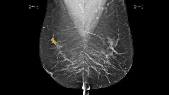
GE HealthCare Breast Cancer AI

GE HealthCare debuts AI suite for early breast cancer detection

MyBreastAI integrates three AI applications designed to read mammograms and measure breast density.

Lunit INSIGHT DBT

Lunit AI-powered 3D breast imaging receives FDA clearance

The product allows radiologists to zoom into breast cancer lesions and gather details on potential malignancy. 

Women in rural areas, especially racial minorities, are less likely to receive a mammogram

A study looking at older Medicare beneficiaries in Texas highlights a need for targeted breast cancer screening outreach.

breast radiologist breast cancer mammography

Ultrasound combined with MRI is better able to identify cancerous lesions in breast tissue

Research out of China found that the pairing proved to be more accurate in identifying cancer in nonmass-like lesions.

breast cancer month ribbon

Patients struggling with addiction, mental health are less likely to be screened for breast cancer

Researchers have shed light on the relationship between breast cancer screening and sociodemographic factors.

Paige Breast Lymph Node Screenshot

FDA grants Paige ‘Breakthrough’ status for breast cancer detection

Paige Lymph Node uses AI to assist pathologists in detecting breast cancer metastases.

breast cancer month ribbon

Most women fail to recognize signs of breast cancer, a third are confused by screening guidelines

Results of a survey from Ohio State University show the public needs a lot more education on breast cancer facts.

black woman breast cancer pink ribbon

Scientists develop MRI-guided ultrasound for burning breast cancer tumors

Scientists from the Huntsman Cancer Institute developed a noninvasive outpatient procedure to treat breast cancer tumors.

Around the web

The nuclear imaging isotope shortage of molybdenum-99 may be over now that the sidelined reactor is restarting. ASNC's president says PET and new SPECT technologies helped cardiac imaging labs better weather the storm.

CMS has more than doubled the CCTA payment rate from $175 to $357.13. The move, expected to have a significant impact on the utilization of cardiac CT, received immediate praise from imaging specialists.

The newly cleared offering, AutoChamber, was designed with opportunistic screening in mind. It can evaluate many different kinds of CT images, including those originally gathered to screen patients for lung cancer. 

Trimed Popup
Trimed Popup