Breast Imaging

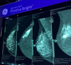
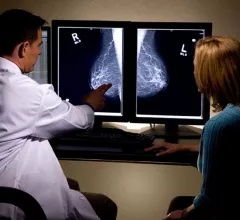
Breast imaging includes imaging modalities used for breast cancer screenings and planning therapy once cancer is detected. Mammography is the primary modality used. Mammogram technology is moving from 2D full-field digital mammography (FFDM) to breast tomosynthesis, or 3D mammography, which helps reduce false positive exams by allowing radiologists to look through the layers of tissue. Overlapping areas of dense breast tissue on 2D mammograms appear similar to cancers and 3D tomo helps determine if suspect areas are cancer or not. About 50% of women have dense breast tissue, which appears white on mammograms, the same as cancers, making diagnosis difficult. Radiologists use the Breast Imaging Reporting and Data System (BI-RADS) scoring system to define the density of breast tissue. Many states now require patients to be notified if they have dense breasts so they understand their mammograms might be suboptimal and they should use supplemental imaging that can see through the dense areas. This includes tomosythesis, breast ultrasound, automated breast ultrasound (ABUS), breast MRI, contrast enhanced mammography and nuclear imaging, including positron emission mammography (PEM).

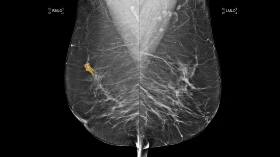
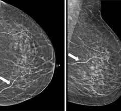
Breast arterial calcifications (BACs) identified on screening mammograms may help identify women who face a heightened risk of developing cardiovascular disease (CVD), according to a new analysis published in Clinical Imaging.

Nearly 25% of women with breast arterial calcifications on mammo develop heart disease

Specifically, women with BACs on their mammogram may be at an increased risk of atherosclerotic cardiovascular disease.

American College of Radiology (ACR) CEO Dana H. Smetherman, MD, MPH, MBA, FACR, explains why opportunistic screening is an important AI imaging technology trend radiology practices should be paying attention.

AI opportunistic screening may have tremendous potential to help patients, ACR CEO says

American College of Radiology leader Dana Smetherman, MD, MBA, discusses the new technology trend and why radiologists should be paying attention. 

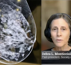
Debra L. Monticciolo, MD, FACR, FSBI, past-president of both the Society of Breast Imaging (SBI), and the American College of Radiology (ACR), explains the current recommendations and a study she did looking at real patient data and projected outcomes using different screening parameters.

The debate over when women should start breast screenings

Debra Monticciolo, MD, who has previously led both the Society of Breast Imaging and the American College of Radiology, discussed how different screening strategies may impact patient outcomes.

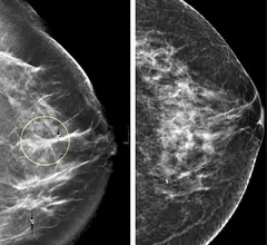
Comparison of a 2D digital mammogram and breast tomosynthesis 3D mammography from UCSF.

90% of breast imaging centers now offer 3D tomosynthesis

Experts in the field say the rising numbers of 3D mammography systems show it is becoming the new standard of care.

ACR CEO outlines top trends in breast imaging

Dana Smetherman, MD, is a diagnostic radiologist who specializes in breast imaging. She spoke to Health Imaging about some key issues that have her attention in 2024 and beyond. 

Thumbnail

LG rolls out new monitors targeted at mammography reads

The new monitor, which was recently approved by the FDA, is intended to improve the process of reading breast MRIs, mammograms and DBT exams.

Breast arterial calcifications (BACs) identified on screening mammograms may help identify women who face a heightened risk of developing cardiovascular disease (CVD), according to a new analysis published in Clinical Imaging.

Experts call for reporting guidelines for breast arterial calcifications identified on mammo

BACs and cardiovascular risk have been a growing topic of conversation in recent years, as numerous studies have indicated there could be a relationship between the two.

Thumbnail

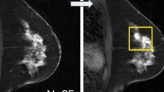
AI tool's supplemental MRI recs help detect 4 times more breast cancers than density measures

What’s more, many of the cancers detected were invasive or multifocal, making their detection more timely. 

Around the web

Positron, a New York-based nuclear imaging company, will now provide Upbeat Cardiology Solutions with advanced PET/CT systems and services. 

The nuclear imaging isotope shortage of molybdenum-99 may be over now that the sidelined reactor is restarting. ASNC's president says PET and new SPECT technologies helped cardiac imaging labs better weather the storm.

CMS has more than doubled the CCTA payment rate from $175 to $357.13. The move, expected to have a significant impact on the utilization of cardiac CT, received immediate praise from imaging specialists.