Nuclear Medicine

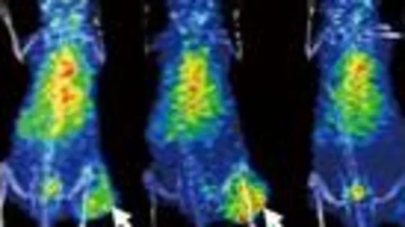
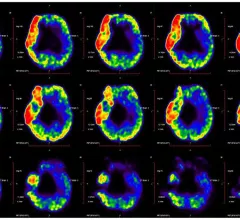
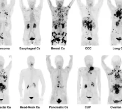
Nuclear medicine (also called molecular imaging) includes positron emission computed tomography (PET) and single photon emission computed tomography (SPECT) imaging. Nuclear imaging is achieved by injecting small amounts of radioactive material (radiopharmaceuticals) into patients before or during their scan. These can use sugars or chemical traits to bond to specific cells. The radioactive material is taken up by cells that consume the sugars. The radiation emitted from inside the body is detected by photon detectors outside the body. Computers take the data to assemble images of the radiation emissions. Nuclear images may appear fuzzy or ghostly rather than the sharper resolution from MRI and CT.  But, it provides metabolic information at a cellular level, showing if there are defects in the function of the heart, areas of very high metabolic activity associated with cancer cells, or areas of inflammation, data not available from other modalities. These noninvasive imaging exams are used to diagnose cancer, heart disease, Alzheimer’s and Parkinson’s disease, bone disorders and other disorders. 

Barry L. Zaret, MD

Veteran cardiologist, viewed by many as the founder of nuclear cardiology, dies at 82

Barry L. Zaret, MD, was an influential cardiologist, accomplished poet and veteran of the United States Air Force. 

October 28, 2022
thalamus

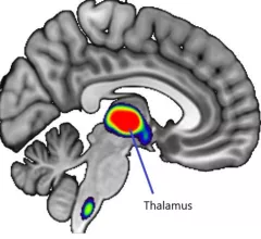
Brain scans reveal single doses of nicotine inhibiting estrogen production in women

On MRI and PET brain scans, women who were exposed to a small dose of nicotine showed altered activity in the thalamus region of the brain.

October 17, 2022
lung CT

CRC patients with these clinical characteristics need more frequent post-op chest imaging

These patients are at greatest risk of developing lung metastases within three months of surgery.

October 17, 2022

PET/CT findings predict post-treatment, radiation-induced hypothyroidism

Radiation-induced hypothyroidism is common yet underdiagnosed, potentially owing to a lack of follow-up consensus in patients treated with radiation therapy for head and neck cancers.

October 13, 2022
Chest pain

FAPI PET/CT findings linked with risk of sudden cardiac death

Experts involved in the research suggested that these findings indicate a potential role for FAPI PET/CT imaging in detecting changes in myocardial fibrosis, noting that it could be more sensitive to earlier changes than standard cardiac MRI.

October 12, 2022
cerebral palsy PET imaging

How PET imaging could benefit children with cerebral palsy

These new findings could help providers differentiate and manage CP associated with different levels of motor impairment, authors of the study indicated.

October 7, 2022
FAPI PET/CT shows promise as a better radiotracer for cancer than 18F-FDG. It also can be used for detection of infection and inflammation.

VIDEO: 6 key advances in molecular imaging technology for PET and SPECT

Munir Ghesani, MD, President of the Society of Nuclear Medicine and Molecular Imaging (SNMMI), system chief of nuclear medicine at Mount Sinai Health, explains recent advances in nuclear imaging technology.

September 30, 2022
lung CT

New lesion measurement assesses treatment responses more accurately than RECIST

Researchers believe the new method of assessing treatment responses could pave the way for developing new cancer therapeutics.

September 30, 2022

Around the web

Automated AI-generated measurements combined with annotated CT images can improve treatment planning and help referring physicians and patients better understand their disease, explained Sarah Jane Rinehart, MD, director of cardiac imaging with Charleston Area Medical Center.

Two advanced algorithms—one for CAC scores and another for segmenting cardiac chamber volumes—outperformed radiologists when assessing low-dose chest CT scans. 

"Gen AI can help tackle repetitive tasks and provide insights into massive datasets, saving valuable time," Thomas Kurian, CEO of Google Cloud, said Tuesday. 

Trimed Popup
Trimed Popup