Computed Tomography

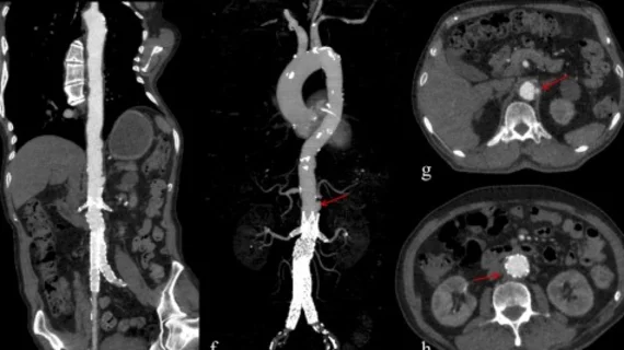
Computed tomography (CT) is a fast and accurate imaging modality often used in emergency settings and trauma imaging. CT scans, with or without (or both) iodinated contrast are frequently used to image the brain, chest, abdomen and pelvis, but also have post-imaging reconstructive capabilities for detailed orthopedic imaging. It is now a standard imaging modality in emergency rooms to quickly assess patients. CT uses a series of X-ray images shot as the gantry rotates around the patient. Computer technology assembles these into into a dataset volume than can be slices on any access, or advanced visualization software can extract specific parts of the anatomy for study. Find more content specific to cardiac CT.
U.S. News & World Report children’s hospitals cardiology heart surgery

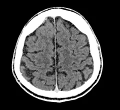
New cancer risk analysis prompts experts to call for 'careful justification' of pediatric head CTs

There is a significant dose-response relationship between radiation incurred during head CT scans and brain cancer in children and young adults, according to a new paper published in The Lancet Oncology.

AI is still one of the key technologies on the floor many radiologists want to learn more about. of the AI. A product rep discussing breast automated detection AI in the crowded Lunit booth at RSNA 2022.

9 technology trends and takeaways from RSNA 2022

Here are some interesting new technologies and key trends from the vast expo floor at the Radiological Society of North America 2022 meeting.

Jorge Soto, MD, chair of the RSNA Annual Meeting Program Planning Committee, chief of radiology, Boston Medical Center, and professor of medicine, Boston University School of Medicine, offers an overview of the trends, hot topics, and innovative research and technology at the Radiological Society of North America (RSNA) 2022 meeting.

VIDEO: Key takeaways from RSNA 2022

Jorge Soto, MD, chair of the RSNA Annual Meeting Program Planning Committee, chief of radiology, Boston Medical Center, offers an overview of the trends, hot topics, research and technology at the Radiological Society of North America (RSNA) 2022 meeting.

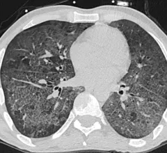
Patient with severe, lung damage from COVID. Images in a 54-year-old man with COVID-19–related acute respiratory distress syndrome and subsequent fibrosis. (A) Axial CT 2 weeks after admission shows diffuse ground-glass opacity (GGO) with reticular abnormality and traction bronchiectasis in right middle lobe, indicating organizing phase of lung injury. (B) Axial CT 6 months after admission shows decreased GGO but extensive traction bronchiectasis and architectural distortion, suggesting fibrosis. RSNA

Lung scarring occurs in up to 11% of patients recovered from COVID, regardless of infection severity

Researchers cannot yet determine whether these changes will progress or resolve in the long term.

Altered mental status and head CT: Study suggests the exam is overutilized at the expense of patients

The meta-analysis included 25 studies and nearly 80,000 patient cases. Experts found that while 94% of those patients with acute atraumatic AMS underwent non-contrast head CT scans, just 11% of those exams yielded positive results. 

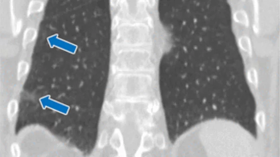
Lung CT of a 42-year-old man with more than 7 days of symptoms. Scans show COVID pneumonia with diffuse ground-glass opacities in close vicinity of visceral pleural surfaces. In addition, a crazy paving pattern is observed. Image courtesy of RSNA. Medical images of COVID. #COVID #SARS-CoV-2

'Crash course' on COVID cases benefits residents less experienced with thoracic CT

Following the course, experts observed nearly perfect agreement between less experienced residents and chest-devoted radiologists.

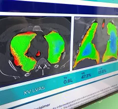
Dynamic lung air flow analysis just using X-ray without any contrast with new technology from 4D Medical.

PHOTO GALLERY: New technology and trends at RSNA 2022

Images from the Radiological Society of North America (RSNA) 2022 annual meeting Nov. 27- Dec. 1 in Chicago. The gallery includes new technologies and a look at sights around the world's largest radiology conference. 

Thumbnail

Experts call for reporting of all incidentally detected coronary calcifications

Experts recently found that reporting of coronary calcium is frequently limited to only moderate to severe cases.

Around the web

Positron, a New York-based nuclear imaging company, will now provide Upbeat Cardiology Solutions with advanced PET/CT systems and services. 

The nuclear imaging isotope shortage of molybdenum-99 may be over now that the sidelined reactor is restarting. ASNC's president says PET and new SPECT technologies helped cardiac imaging labs better weather the storm.

CMS has more than doubled the CCTA payment rate from $175 to $357.13. The move, expected to have a significant impact on the utilization of cardiac CT, received immediate praise from imaging specialists.

Trimed Popup
Trimed Popup