Computed Tomography

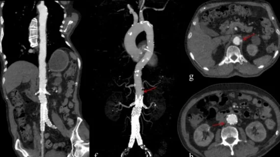
Computed tomography (CT) is a fast and accurate imaging modality often used in emergency settings and trauma imaging. CT scans, with or without (or both) iodinated contrast are frequently used to image the brain, chest, abdomen and pelvis, but also have post-imaging reconstructive capabilities for detailed orthopedic imaging. It is now a standard imaging modality in emergency rooms to quickly assess patients. CT uses a series of X-ray images shot as the gantry rotates around the patient. Computer technology assembles these into into a dataset volume than can be slices on any access, or advanced visualization software can extract specific parts of the anatomy for study. Find more content specific to cardiac CT.
Leslee Shaw, PhD, MSCCT, FACC, MASNC, FAHA, director of the Blavatnik Family Women's Health Research Institute, a professor of medicine at the Icahn School of Medicine at Mount Sinai, and a former president of both SCCT and ASNC. She explains the sex differences in cardiovascular imaging presentations in women versus men.

VIDEO: CT can play a role in identifying women's differences in cardiovascular presentations

Leslee Shaw, PhD, director of the Blavatnik Family Women's Health Research Institute, a professor of medicine at the Icahn School of Medicine at Mount Sinai, and a former president of both SCCT and ASNC, explains the sex differences in cardiovascular imaging presentations in women versus men. 

Ricardo Cury, MD, MBA, MSCCT, chairman of radiology, direct of cardiac imaging, Baptist Health South Florida and Miami Cardiac and Vascular Institute, discusses the new CAD-RADS 2.0 cardiac imaging reporting criteria at the 2022 SCCT meeting. Interview with Radiology Business Editor Dave Fornell.

VIDEO: What is new with CAD-RADS 2.0 cardiac imaging reporting?

Ricardo Cury, MD, chairman of radiology and director of cardiac imaging, Baptist Health South Florida and Miami Cardiac and Vascular Institute, discusses the new CAD-RADS 2.0 cardiac imaging reporting criteria.

AI-generated coronary tree from a patient's CT scan showing a color code of areas of interest for plaque burden from the Cleerly software shown at SCCT 2022.

VIDEO: The role of AI in cardiac imaging

Ed Nicol, MD, president-elect of the Society of Cardiovascular Computed Tomography, provided us with an exclusive look at how AI is expected to change cardiac imaging.

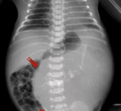
Triplet pregnancy fetus in fetu: A rare case report

While FIF is an extremely rare condition—occurring in about one in 500,000 births—it is even more rare in triplet pregnancies.

Thumbnail

Digitized informed consent increases detection of critical 'red flags' prior to imaging exams

Digitized informed patient consent prior to contrast-enhanced CT exams has significant advantages over traditional paper consent forms, but it also has the unfortunate effect of leaving some of the most vulnerable patients behind.  

long covid lung CT

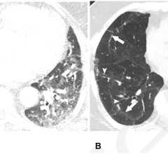
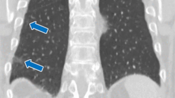
More than half of long COVID patients have abnormalities on chest imaging

Nodular patterns were the most commonly visualized abnormality, although several others were observed.

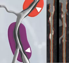
Left, HeartFlow's RoadMap analysis enables cardiac CT readers to identify stenoses in the major coronary arteries. The AI provides visualization and quantification of the location and severity of anatomic narrowings. Right image, HeartFlow's Plaque Analysis AI algorithm automates assessment of coronary plaque characteristics and volume on CCTA exams to greatly reduce the time it takes to manually assess and quantify these features.

HeartFlow gains FDA clearance for 2 new AI-powered imaging assessments

The solutions, Plaque Analysis and RoadMap Analysis, both use coronary CT angiography to provide clinicians with a noninvasive look at patients who present with coronary artery disease and face a heightened myocardial infarction risk.

Thumbnail

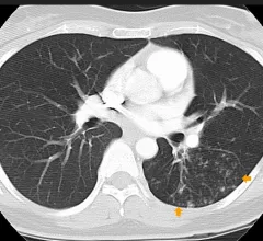
CRC patients with these clinical characteristics need more frequent post-op chest imaging

These patients are at greatest risk of developing lung metastases within three months of surgery.

Around the web

Positron, a New York-based nuclear imaging company, will now provide Upbeat Cardiology Solutions with advanced PET/CT systems and services. 

The nuclear imaging isotope shortage of molybdenum-99 may be over now that the sidelined reactor is restarting. ASNC's president says PET and new SPECT technologies helped cardiac imaging labs better weather the storm.

CMS has more than doubled the CCTA payment rate from $175 to $357.13. The move, expected to have a significant impact on the utilization of cardiac CT, received immediate praise from imaging specialists.

Trimed Popup
Trimed Popup