Magnetic Resonance Imaging

Magnetic resonance imaging (MRI) is the gold standard imaging modality for soft tissues. It produces detail cross-sectional images of soft tissue and bone anatomy, including muscles, tendons, ligaments, brain and organs, without the use of ionizing radiation. In addition to orthopedic imaging, MRI is also used for heart, brain and breast. MRI uses gadolinium contrast in many exams to highlight tissues and blood vessels, which enhances images and offers better diagnostic quality. It can also be used in conjunction with PET scans. How does MRI work? MR creates images by using powerful magnets to polarize hydrogen atoms in water (the body is made of of more than 80% water) so they face in one direction. A radiofrequency pulse is then used to ping these atoms, causing them to wobble, or resonate. The MRI coils detect this and computers can assemble images from the signals. Basic MRI scans will focus on the resonance of fat and water in two different sequences, which highlight and contrast different features in the anatomy.
doctor looking at CLTI patient's leg

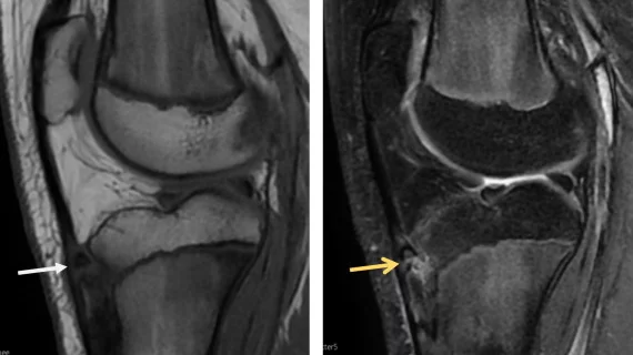
New MRI technology improves imaging of metallic joint replacements

The technology is so promising that it is being integrated into GE Healthcare’s MRI scanners so that providers can conduct more thorough evaluations of how these implants hold up over time.

gaming keyboard

How gaming addiction affects brain activity, according to new MRI data

Over time, the brain's reward system may become less responsive to standard rewards in individuals with gaming addiction.

Thumbnail

How opioid use disorder affects brain size and function

The CDC estimates that over 6 million people in the U.S. struggle with OUD, and prior research has indicated that only 20% receive treatment for the disorder.

Radiomics model links brain MRI features with neuropsychological changes in Crohn's disease

Researchers hope their findings can help pave the way for more targeted therapies for individuals with Crohn's disease.

money court scale of justice ruling legal settlement

Nearly $800,000 settlement offered to family of man who died during MRI exam

The patient, who was being cared for in the ICU, was not accompanied or monitored by nursing staff during his exam, despite being sedated.

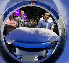
RSNA attendees through bore of the new release Philips CT 5300 scanner at RSNA 2024. Three new CT systems were released at RSNA that included numerous AI enhancements. Photo by Dave Fornell. #RSNA #RSNA24 #RSNA2024

Key takeaways from RSNA 2024 for AI, CT, MRI and the radiology staffing shortage

Several key trends were evident at the Radiological Society of North America 2024 meeting, including new CT and MR technology and evolving adoption of artificial intelligence.

Thumbnail

Some soccer players are at risk of brain damage similar to CTE

American football isn’t the only contact sport that has medical experts concerned with the brain health of athletes. New evidence suggests certain soccer players may be at heightened risk of long-term brain abnormalities, too. 

Thumbnail

GBCA dose reduced by up to 80% with help from deep learning-based image reconstruction

Although GBCAs are largely considered safe, there are concerns about how gadolinium retention could affect patients who require repeated imaging.

Around the web

To fully leverage today's radiology IT systems, standardization is a necessity. Steve Rankin, chief strategy officer for Enlitic, explains how artificial intelligence can help.

RBMA President Peter Moffatt discusses declining reimbursement rates, recruiting challenges and the role of artificial intelligence in transforming the industry.

Deepak Bhatt, MD, director of the Mount Sinai Fuster Heart Hospital and principal investigator of the TRANSFORM trial, explains an emerging technique for cardiac screening: combining coronary CT angiography with artificial intelligence for plaque analysis to create an approach similar to mammography.