Magnetic Resonance Imaging

Magnetic resonance imaging (MRI) is the gold standard imaging modality for soft tissues. It produces detail cross-sectional images of soft tissue and bone anatomy, including muscles, tendons, ligaments, brain and organs, without the use of ionizing radiation. In addition to orthopedic imaging, MRI is also used for heart, brain and breast. MRI uses gadolinium contrast in many exams to highlight tissues and blood vessels, which enhances images and offers better diagnostic quality. It can also be used in conjunction with PET scans. How does MRI work? MR creates images by using powerful magnets to polarize hydrogen atoms in water (the body is made of of more than 80% water) so they face in one direction. A radiofrequency pulse is then used to ping these atoms, causing them to wobble, or resonate. The MRI coils detect this and computers can assemble images from the signals. Basic MRI scans will focus on the resonance of fat and water in two different sequences, which highlight and contrast different features in the anatomy.

Female Medical Research Scientist Working with Brain Scans

New findings prompt experts to call for routine brain imaging of certain breast cancer patients

New research suggests more women with late-stage breast cancer may develop brain metastases than previously believed. 

cannabis use disorder marijuana joint weed smoking

MRI shows 'long-lasting' impact of cannabis use on brain regions linked to memory

These findings should serve as a reminder that marijuana, which is often touted as a safer and more natural alternative to alcohol, can cause harm. 

MRI technique analyzes composition of fat surrounding heart.

New MRI technique spots signs of heart disease before symptoms emerge

Researchers with the University of Virginia Arlington are working to develop a new way of analyzing fat content around the heart. 

Thumbnail

Alzheimer's and Parkinson's cause similar cognitive symptoms, but show opposing patterns on MRI

This new information on the pathogenesis of both conditions could be useful for the development of interventions targeted at slowing cognitive decline. 

COVID-19 coronavirus mask smell

Chemical alterations in the brain hint at whether loss of smell will return post-COVID

Researchers have identified what they believe could serve as imaging biomarkers that predict recovery from anosmia after contracting the virus. 

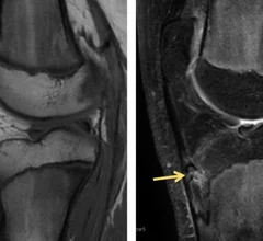
MRI of knee with Osgood-Schlatter Disease

MRI scoring system simplifies diagnosis of common adolescent ortho issue

The condition can worsen with physical activity, which is why an accurate and timely diagnosis is important for healthy growth. 

birth control the pill cardiovascular disease blood clots side effects

Researcher scans herself 75 times to study effects of birth control on brain health

The postdoctoral research fellow hopes her research will provide greater insight into how the use of oral birth control impacts physical and mental health.

Thumbnail

Brain changes related to Huntington's evident on imaging decades before symptoms emerge

Experts hope their findings offer valuable insights that one day lead to improved interventions aimed at preserving brain function in patients with Huntington's. 

Around the web

GE HealthCare designed the new-look Revolution Vibe CT scanner to help hospitals and health systems embrace CCTA and improve overall efficiency.

Clinicians have been using HeartSee to diagnose and treat coronary artery disease since the technology first debuted back in 2018. These latest updates, set to roll out to existing users, are designed to improve diagnostic performance and user access.

The cardiac technologies clinicians use for CVD evaluations have changed significantly in recent years, according to a new analysis of CMS data. While some modalities are on the rise, others are being utilized much less than ever before.