Magnetic Resonance Imaging

Magnetic resonance imaging (MRI) is the gold standard imaging modality for soft tissues. It produces detail cross-sectional images of soft tissue and bone anatomy, including muscles, tendons, ligaments, brain and organs, without the use of ionizing radiation. In addition to orthopedic imaging, MRI is also used for heart, brain and breast. MRI uses gadolinium contrast in many exams to highlight tissues and blood vessels, which enhances images and offers better diagnostic quality. It can also be used in conjunction with PET scans. How does MRI work? MR creates images by using powerful magnets to polarize hydrogen atoms in water (the body is made of of more than 80% water) so they face in one direction. A radiofrequency pulse is then used to ping these atoms, causing them to wobble, or resonate. The MRI coils detect this and computers can assemble images from the signals. Basic MRI scans will focus on the resonance of fat and water in two different sequences, which highlight and contrast different features in the anatomy.

Thumbnail

MRI findings linked to cognitive issues in patients with long COVID 2 years post-infection

The CDC estimates that just under 7% of adults in the United States continue to struggle with symptoms of long COVID.

Thumbnail

MRI study reveals never-before-seen changes in the brain during pregnancy

While prior imaging studies have compared changes that occur in the brain before pregnancy and after childbirth, this latest analysis is the first to span multiple time points during the full gestational period.

Cardiac PET on the rise among U.S. cardiologists

SPECT is still the most common modality used to evaluate CAD patients, but cardiac PET is gaining more and more momentum.

Thumbnail

Polaris Dawn astronauts will undergo brain MRIs upon landing

The scans will be conducted earlier than any other post-flight imaging of astronauts on record. 

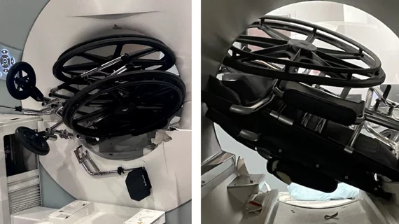
MRI accident prompts questions on safety

Yet another MRI 'freak accident' is making headlines months after it took place

When it happened, news of the incident did not circulate, and whether it was reported to any governing body remains unclear.

MRI image of a fetus inside the mother on an MRI scan to assess brain development. Image courtesy of RSNA.

Fetal MRI safe on 3T scanners, new analysis shows

MRI exams are crucial for investigating fetal abnormalities, but past concerns have been expressed about whether the modality could affect intrauterine growth. 

Thumbnail

MRI study questions links between autism and brain connectivity

Prior studies have hypothesized that reduced functional connectivity in the amygdala was more common among people on the spectrum.

Thumbnail

Some people may be predisposed to depression, MRI study shows

Specific brain connectivity patterns evident during childhood might make people more vulnerable to developing psychiatric disorders.

Around the web

GE HealthCare's flurpiridaz, the PET radiotracer that recently received FDA approval, offers several key benefits over SPECT. Jamshid Maddahi, MD, discussed the details in an exclusive interview. 

Ultrafast MCE could go on to become a go-to treatment option for obstructive coronary artery disease, according to the authors of a new first-in-human clinical study.

Elucid's PlaqueIQ was trained to turn CCTA images into interactive 3D reports that help physicians visualize the presence of atherosclerosis.

Trimed Popup
Trimed Popup