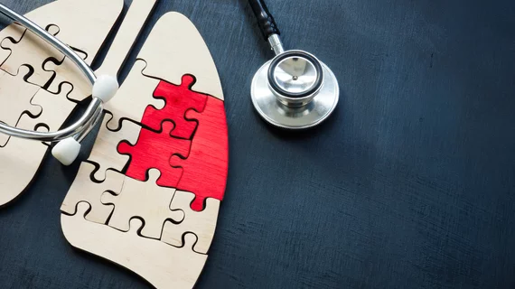
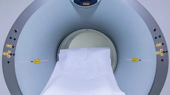
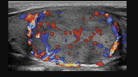
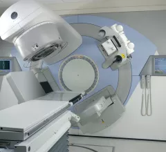
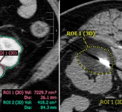
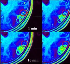
Oncology Imaging

Medical imaging has become integral to cancer care, assessing the stage and location of cancerous tumors. By utilizing powerful imaging modalities including CT, MRI, MRA and PET/CT, oncology imaging radiologists are able to assist referring physicians in the detection and diagnosis of cancer.

FAPI PET improves staging and clinical management for a significant portion of pancreatic cancers

Fibroblast activation protein inhibitor PET imaging may provide more accurate insight into the spread of pancreatic cancer than more commonly used 18F-FDG PET/CT.

Thumbnail

Reducing prostate cancer treatment dosage alleviates downsides while remaining effective

Reducing certain treatments by around 25% and 50% still achieves promising PSA response rates, but reduces some of the unwanted side effects of therapy, like dry mouth. 

Thumbnail

Researchers score $2M to examine side effects of radiation therapy in prostate cancer patients

The grant will fund a team at the UCLA Health Jonsson Comprehensive Cancer Center working to identify genetic biomarkers capable of predicting RT side effects.

renal cryoablation volumes

How much is too much when it comes to cryoablation volumes in solitary kidneys?

A paper in the Journal of Vascular and Interventional Radiology offers new guidance on how ablation volumes affect renal function in solitary kidneys and highlights ranges that pose the least risk to patients. 

Thumbnail

Fluorinated contrast agents show 'strong potential' for MRI theranostics

Experts have developed a first-of-its-kind fluorinated contrast agent they believe will provide more detailed MR imaging and aid in the treatment of certain cancers. 

colorectal cancer colon CTC CRC colonoscopy

Radiology's role in colorectal cancer care set to grow following 'groundbreaking' trial results

The results were so conclusive that the trial was halted at halftime for having met predefined stopping rules. 

Lung cancer screening CT image in a 66-year-old male patient shows a sessile nodule with internal air in the left mainstem to left upper lobe bronchus (arrow) with a mean diameter of 10 mm. The nodule was assigned as Lung-RADS category 4A in the clinical report. (B) Follow-up CT image shows the lesion is resolved. Image courtesy of RSNA

Lung-RADS update helps limit false-positive results, unnecessary procedures

Using the latest version was associated with improved diagnostic accuracy, researchers wrote in a new analysis. 

time-delayed MRI phases improve brain tumor visualization

Using multiple MRI phases after contrast injection significantly improves visualization of brain tumors

Researchers are proposing making additional phases beyond the 10-minute mark mandatory when analyzing brain metastases.

Around the web

Harvard’s David A. Rosman, MD, MBA, explains how moving imaging outside of hospitals could save billions of dollars for U.S. healthcare.

Back in September, the FDA approved GE HealthCare’s new PET radiotracer, flurpiridaz F-18, for patients with known or suspected CAD. It is seen by many in the industry as a major step forward in patient care. 

After three years of intermittent shortages of nuclear imaging tracer technetium-99m pyrophosphate, there are no signs of the shortage abating.