Computed Tomography

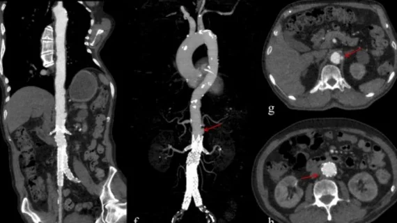
Computed tomography (CT) is a fast and accurate imaging modality often used in emergency settings and trauma imaging. CT scans, with or without (or both) iodinated contrast are frequently used to image the brain, chest, abdomen and pelvis, but also have post-imaging reconstructive capabilities for detailed orthopedic imaging. It is now a standard imaging modality in emergency rooms to quickly assess patients. CT uses a series of X-ray images shot as the gantry rotates around the patient. Computer technology assembles these into into a dataset volume than can be slices on any access, or advanced visualization software can extract specific parts of the anatomy for study. Find more content specific to cardiac CT.

Imaging surveillance program detects pancreatic cancer in its earliest stages

Individuals at high risk of pancreatic cancer benefit from annual imaging and have decreased mortality rates compared to those who forgo preventive screening.  

Thumbnail

Content-based AI system decreases read times by 31% while increasing accuracy

Using CBIRS decreased interpretation times despite the radiologists who utilized them having to review the additional information the system provided.

Thumbnail

Commercial PE-detecting algorithm identifies incidental clots on CT

Experts involved in the study, which analyzed more than 3,000 CT scans, suggested that there could be a future role for the algorithm to assist radiologists in busy settings.

Aidoc announces strategic hospital partnership that will advance AI in clinical settings

The software will be used for the triage and notification of intracranial hemorrhage (ICH), pulmonary embolism (PE) and c-spine fractures identified on CT imaging.

Thumbnail

CT scans set to take flight in new project that aims to integrate imaging into air ambulances

The Norwegian Air Ambulance Foundation is using a newly acquired five-bladed H145 helicopter to develop and test the technology and hopes to have a CT scanner implemented into it in the next four to five years. 

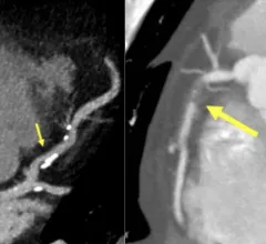
Examples of new plaque reporting in the CAD-RADS 2.0 document. Left, an example from CAD-RADS 2 / P2 plaque burden with mild coronary stenosis (25-49%). Right, example of a CAD-RADS 5/ P3, with a focal, non-calcified occlusion of the proximal RCA (arrow) and severe amount of plaque (P3). #CADRADS #YesCCT #CTA #CCTA

New CAD-RADS 2.0 reporting for coronary CTA offers patient management recommendations

The document includes updated classification to established a framework for stenosis, plaque burden and plaque modifiers, including assessment of CT-FFR or myocardial CT perfusion.

Contrast shortage update: Supplies could continue to lag until September

GE previously indicated that the iodinated contrast shortage would resolve by the end of June.

Thumbnail

Reducing image-guided lung biopsy complications

Experts observed significantly reduced pneumothorax and pulmonary hemorrhage rates of 8% and 6% when utilizing the method.

Around the web

Positron, a New York-based nuclear imaging company, will now provide Upbeat Cardiology Solutions with advanced PET/CT systems and services. 

The nuclear imaging isotope shortage of molybdenum-99 may be over now that the sidelined reactor is restarting. ASNC's president says PET and new SPECT technologies helped cardiac imaging labs better weather the storm.

CMS has more than doubled the CCTA payment rate from $175 to $357.13. The move, expected to have a significant impact on the utilization of cardiac CT, received immediate praise from imaging specialists.