Computed Tomography

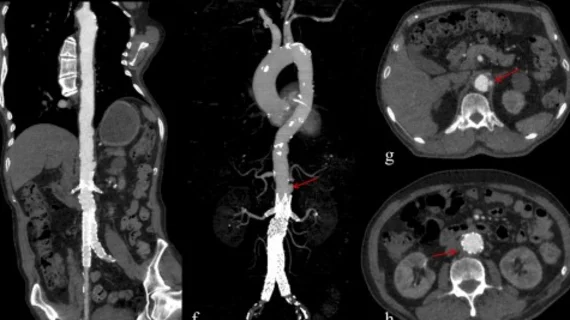
Computed tomography (CT) is a fast and accurate imaging modality often used in emergency settings and trauma imaging. CT scans, with or without (or both) iodinated contrast are frequently used to image the brain, chest, abdomen and pelvis, but also have post-imaging reconstructive capabilities for detailed orthopedic imaging. It is now a standard imaging modality in emergency rooms to quickly assess patients. CT uses a series of X-ray images shot as the gantry rotates around the patient. Computer technology assembles these into into a dataset volume than can be slices on any access, or advanced visualization software can extract specific parts of the anatomy for study. Find more content specific to cardiac CT.
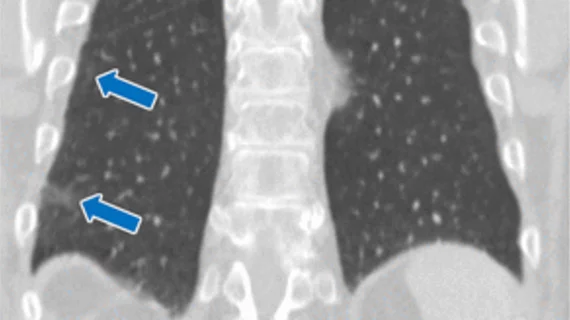
lung cancer pulmonary nodule

How effective are guidelines for managing incidental pulmonary nodules spotted on CT?

The authors of the study hypothesized that they would find malalignments due to the nature of how the 2017 Fleischner Society guidelines, in that they do not pertain to incidentally detected nodules.

pancreas lesions on CT

DL model's pancreatic lesion detection in line with that of board-certified radiologists

“Our approach has the potential to facilitate timely diagnoses and management of pancreatic lesions encountered in routine clinical practice,” experts involved in the study said.

Fully automated CT body composition analysis predicts survival for CRC patients

A fully automated body composition analysis derived from CT imaging can be a valuable pretreatment tool for patients with colorectal cancer. 

non-small cell lung cancer tumor segmentation

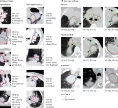
Algorithm reduces NSCLC tumor segmentation times by 65%

In a close collaboration with radiation oncologists, experts trained their model on the CT lung images of 787 patients and tested it on the scans of more than 1,300 patients from external datasets.

Thumbnail

Why do many of America's most at-risk veterans decline annual CT lung cancer screening?

The new analysis included 43,000 eligible U.S. veterans, 32% of whom were observed to have declined annual CT screening for lung cancer.

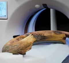
woolly mammoth tusk CT

A mammoth accomplishment: CT captures images of an entire ancient tusk

Prior attempts at imaging large fossils such as mammoth tusks failed to capture the full artifact with just one scan, instead requiring multiple partial scans that were subsequently pieced together.

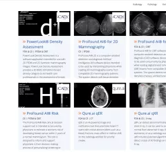
Example of an artificial intelligence (AI) app store on the Sectra website, where Sectra PACS users can select the AI algorithms they want that are already integrated into the Sectra System. Other vendors have followed a similar approach to AI developed by many smaller vendors they partner with.

VIDEO: Development of AI app stores to enable easier access

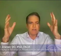
Keith J. Dreyer, DO, PhD, FACR, American College of Radiology (ACR) Data Science Institute Chief Science Officer, explains how radiology vendors have developed AI app stores to make it easier to access new FDA cleared AI algorithms.
 

Keith J. Dreyer, DO, PhD, FACR, American College of Radiology (ACR) Data Science Institute Chief Science Officer, explains artificial intelligence (AI) for radiology. Dreyer also holds the positions of vice chairman of radiology at Massachusetts General Hospital, chief data science and information officer for the departments of radiology for both Massachusetts General Hospital and Brigham and Women's Hospital.

VIDEO: Where will radiology AI be in 5 years?

Keith J. Dreyer, DO, PhD, FACR, American College of Radiology (ACR) Data Science Institute Chief Science Officer, explains 5 developments to watch for in radiology artificial intelligence (AI).

Around the web

Positron, a New York-based nuclear imaging company, will now provide Upbeat Cardiology Solutions with advanced PET/CT systems and services. 

The nuclear imaging isotope shortage of molybdenum-99 may be over now that the sidelined reactor is restarting. ASNC's president says PET and new SPECT technologies helped cardiac imaging labs better weather the storm.

CMS has more than doubled the CCTA payment rate from $175 to $357.13. The move, expected to have a significant impact on the utilization of cardiac CT, received immediate praise from imaging specialists.