Computed Tomography

Computed tomography (CT) is a fast and accurate imaging modality often used in emergency settings and trauma imaging. CT scans, with or without (or both) iodinated contrast are frequently used to image the brain, chest, abdomen and pelvis, but also have post-imaging reconstructive capabilities for detailed orthopedic imaging. It is now a standard imaging modality in emergency rooms to quickly assess patients. CT uses a series of X-ray images shot as the gantry rotates around the patient. Computer technology assembles these into into a dataset volume than can be slices on any access, or advanced visualization software can extract specific parts of the anatomy for study. Find more content specific to cardiac CT.
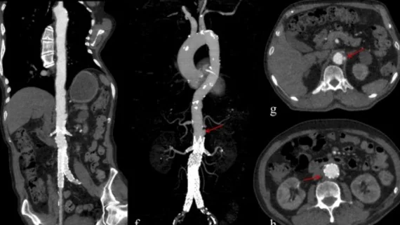
Vascular CT beyond the coronaries includes imaging for aortic aneurism, aortic dissection, SCAD and fibromuscular dysplasia. #SCCT #SCCT2022 #SCAD #yescct

VIDEO: Vascular imaging applications for CT beyond the heart

Society of Cardiovascular Computed Tomography (SCCT) President Brian Ghoshhajra, MD, explains the vascular imaging sessions at SCCT 2022 that went beyond the usual coronaries and structural heart evaluations.

coronary CTA

Deep learning, subtraction technique ideal for evaluating stent re-stenosis on coronary CTA

Detecting in-stent restenosis via coronary CTA with hybrid iterative reconstruction has historically been an exercise in avoiding false positives.

Thumbnail

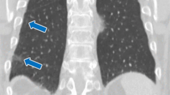
These CT findings increase risk of thromboembolic events for patients with COVID pneumonia

Authors of the new paper noted that radiologists should be highly suspicious of pulmonary embolism and DVT when they encounter these findings on chest CT exams of COVID patients.

COVID-19 medical imaging examples of various clinical presentations. SARS-CoV-2 clinical imaging presentations.

PHOTO GALLERY: What does COVID-19 look like on medical imaging?

This image gallery shows what the various clinical presentations associated with the COVID-19 virus that have been documented during the coronavirus pandemic.

Less experienced radiologists benefit from deep learning models when scouting for intracranial aneurysms

Deep learning models can increase reader accuracy while simultaneously decreasing interpretation times when evaluating imaging for intracranial aneurysms.

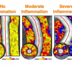
Fat attenuation index (FAI) CT imaging of coronary artery fat can show inflammation and can help pin-point vulnerable plaques, or show the reversal of inflammation due to drug therapies. The technology is being developed by the vendor Caristo, which has European CE mark and the company is seeking FDA clearance. #SCCT #SCCT2022

VIDEO: New Technologies in Cardiac CT Imaging

Ron Blankstein, MD, Brigham and Women's Hospital, explains recent advances in coronary computed tomography angiography (CCTA) technology. 

radiology reporting EHR health record CDS AUC

EHR-based solutions to the iodinated contrast shortage reduce usage by 12%

This week in AJR, experts from a large, multisite health system detailed their efforts to preserve contrast supplies by implementing electronic health record (EHR) order entry-based interventions.

Cardiac CT now in the spotlight thanks to chest pain guidelines

With the elevation of cardiac CT to a 1A indication in the 2021 Chest Pain Guidelines, there has been a large amount of interest in starting coronary artery CT angiography (CCTA) programs. 

Around the web

Positron, a New York-based nuclear imaging company, will now provide Upbeat Cardiology Solutions with advanced PET/CT systems and services. 

The nuclear imaging isotope shortage of molybdenum-99 may be over now that the sidelined reactor is restarting. ASNC's president says PET and new SPECT technologies helped cardiac imaging labs better weather the storm.

CMS has more than doubled the CCTA payment rate from $175 to $357.13. The move, expected to have a significant impact on the utilization of cardiac CT, received immediate praise from imaging specialists.