Computed Tomography

Computed tomography (CT) is a fast and accurate imaging modality often used in emergency settings and trauma imaging. CT scans, with or without (or both) iodinated contrast are frequently used to image the brain, chest, abdomen and pelvis, but also have post-imaging reconstructive capabilities for detailed orthopedic imaging. It is now a standard imaging modality in emergency rooms to quickly assess patients. CT uses a series of X-ray images shot as the gantry rotates around the patient. Computer technology assembles these into into a dataset volume than can be slices on any access, or advanced visualization software can extract specific parts of the anatomy for study. Find more content specific to cardiac CT.
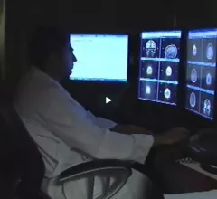
Arl Van Moore, MD, American College of Radiology (ACR) delegate to the American Medical Association (AMA) House of Delegates, former ACR president, chairman of the ACR Board and former CEO of Strategic Radiology, discusses two radiology related policy decisions at the AMA 2022 meeting. These included a AMA policy on the iodine contrast shortage and AUC CDS. #AMA #AMA22 #AMAmtg #AMA175 #ACR

VIDEO: Radiology takeaways from the 2022 AMA meeting

Arl Van Moore, MD, American College of Radiology (ACR) delegate to the American Medical Association House of Delegates, former ACR president, chairman of the ACR Board and former CEO of Strategic Radiology, discusses radiology-related policy decisions at the AMA 2022 meeting.

Thumbnail

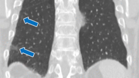
25% of patients undergoing transthoracic needle biopsy experience complications

Higher complication rates were observed in inpatient settings and in patients with COPD. Also associated with increased adverse events were prior lung cancer screening and the use of oral anticoagulants or antiplatelets.

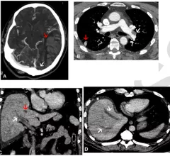
Multimodality imaging turns up serial thromboses following AstraZeneca COVID vaccination

Whole-body imaging reveals clinically undetectable blood clots in patients after their first dose of AstraZeneca's COVID vaccine.

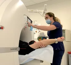
Siemens debuts new, cleared SPECT/CT model

Siemens Healthineers splashed an FDA-approved SPECT/CT system June 12 at the 2022 annual meeting of the Society of Nuclear Medicine & Nuclear Imaging in Vancouver, B.C.

Thumbnail

Radiologists can reclaim an hour every day with AI assistance

The AI software assisted in various tasks, such as segmenting, labeling and measuring normal structures, providing an automated analysis of pulmonary, cardiac and musculoskeletal findings.

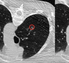
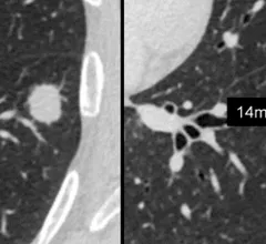
CT scan showing lung cancer nodules with measurements of each nodule to track growth or regression from treatment. Image courtesy of RSNA

Downgrading lung nodules at 3-month follow-up 'may be problematic'

A new analysis suggests that follow-up imaging protocols for certain lung nodules identified via CT may need to be revisited. 

There have been a big increase in interest in cardiac CT following the publication of the 2021 ACC Chest Pain Evaluation Guidelines, which raised CT angiography (CTA) to a 1A level of evidence. This has prompted many centers to consider creating CTA programs.

VIDEO: Cardiac CT training requirements for radiologists and technologists

Brian Ghoshhajra, MD, division chief, cardiovascular imaging, Massachusetts General Hospital, explains what specialized training is needed to perform coronary computed tomography angiography (CCTA) as interest rapidly rises in this field. 

Predicting healthcare utilization in COPD patients using CT and machine learning

Combining CT lung measurements with machine learning models to predict prognosis in COPD patients could help to lessen their reliance on emergency services. 

Around the web

Positron, a New York-based nuclear imaging company, will now provide Upbeat Cardiology Solutions with advanced PET/CT systems and services. 

The nuclear imaging isotope shortage of molybdenum-99 may be over now that the sidelined reactor is restarting. ASNC's president says PET and new SPECT technologies helped cardiac imaging labs better weather the storm.

CMS has more than doubled the CCTA payment rate from $175 to $357.13. The move, expected to have a significant impact on the utilization of cardiac CT, received immediate praise from imaging specialists.