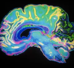
Magnetic Resonance Imaging

Magnetic resonance imaging (MRI) is the gold standard imaging modality for soft tissues. It produces detail cross-sectional images of soft tissue and bone anatomy, including muscles, tendons, ligaments, brain and organs, without the use of ionizing radiation. In addition to orthopedic imaging, MRI is also used for heart, brain and breast. MRI uses gadolinium contrast in many exams to highlight tissues and blood vessels, which enhances images and offers better diagnostic quality. It can also be used in conjunction with PET scans. How does MRI work? MR creates images by using powerful magnets to polarize hydrogen atoms in water (the body is made of of more than 80% water) so they face in one direction. A radiofrequency pulse is then used to ping these atoms, causing them to wobble, or resonate. The MRI coils detect this and computers can assemble images from the signals. Basic MRI scans will focus on the resonance of fat and water in two different sequences, which highlight and contrast different features in the anatomy.

prostate cancer PSA

MRI-based prostate cancer screening paired with new blood test can reduce deaths by up to 9%

The novel approach can also reduce the number of MR scans performed over a lifetime by 60%, experts reported in European Urology.

Thumbnail

Arizona State University home to new 9.4T cryogen-free MRI system

The high-field scanner takes advantage of an innovative dry technology that removes the need for liquid helium and liquid nitrogen required by most MRI systems.

LEGO's miniature MRI models help comfort nervous kids before exams

The company hopes the model can make scans "more playful and less scary" for pediatric patients.

Boston researchers hope PET-MRI brain scans will shed light on 'long-COVID' symptoms

The team will be recruiting patients who are still experiencing extreme fatigue and difficulty concentrating after recovering from the virus.

breast cancer mammography mammogram

Quality improvements reduce biopsy wait times for patients with suspicious breast MRIs

The initiative led to a 30% increase in the number of patients who required fewer visits before receiving a diagnosis, University of Michigan Health System researchers reported.

Thumbnail

Cleveland Clinic to utilize brain MRIs in massive 20 year neurological disease study

“Our hope is to change the course of neurodegeneration, with the long-term goal of curing diseases in their earliest stages, years before symptoms are even seen,” a doctor involved in the study said.

Thumbnail

MRI-based measurements can predict urinary continence after prostate surgery

Radiologists should complete specific training to understand these measurements before using membranous urethra length as a predictor of postoperative continence, University of Michigan researchers suggested.

Thumbnail

6 tips for making MRI exams more autism-friendly

Clinicians should focus on ensuring exams are more compatible with autism patients rather than making such individuals more MR-compatible.

Around the web

The nuclear imaging isotope shortage of molybdenum-99 may be over now that the sidelined reactor is restarting. ASNC's president says PET and new SPECT technologies helped cardiac imaging labs better weather the storm.

CMS has more than doubled the CCTA payment rate from $175 to $357.13. The move, expected to have a significant impact on the utilization of cardiac CT, received immediate praise from imaging specialists.

The newly cleared offering, AutoChamber, was designed with opportunistic screening in mind. It can evaluate many different kinds of CT images, including those originally gathered to screen patients for lung cancer. 

Trimed Popup
Trimed Popup