Magnetic Resonance Imaging

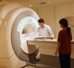
Magnetic resonance imaging (MRI) is the gold standard imaging modality for soft tissues. It produces detail cross-sectional images of soft tissue and bone anatomy, including muscles, tendons, ligaments, brain and organs, without the use of ionizing radiation. In addition to orthopedic imaging, MRI is also used for heart, brain and breast. MRI uses gadolinium contrast in many exams to highlight tissues and blood vessels, which enhances images and offers better diagnostic quality. It can also be used in conjunction with PET scans. How does MRI work? MR creates images by using powerful magnets to polarize hydrogen atoms in water (the body is made of of more than 80% water) so they face in one direction. A radiofrequency pulse is then used to ping these atoms, causing them to wobble, or resonate. The MRI coils detect this and computers can assemble images from the signals. Basic MRI scans will focus on the resonance of fat and water in two different sequences, which highlight and contrast different features in the anatomy.

Thumbnail

Abbreviated MRI outperforms DBT for detecting secondary breast cancer

“Our study results suggest that AB-MRI could potentially be sufficient for post-treatment surveillance,” experts suggested Thursday in Academic Radiology.

Thumbnail

Researchers are developing a modified MRI protocol to help treat brain hemorrhages

“We are studying whether MRI can provide more complete information which could alter the clinical management of patients with hemorrhagic stroke," an expert involved in the trial explained.

Thumbnail

Doctors suggest performing whole-spine MRI scans for suspected child abuse victims

Replacing cervical spine exams with whole-spine MRIs can reveal "major findings," investigators explained recently.

Thumbnail

Combining functional MRI scans with eye tests could help stroke survivors recover lost vision

Identifying the exact location of the damage in a patient's visual pathway may enable doctors to develop unique rehabilitation methods,  experts explained in Frontiers in Neuroscience.

Thumbnail

Doctors pilot mobile stroke unit equipped with low-cost MRI machine

Medical University of South Carolina experts believe they are the first to place a portable magnetic resonance imaging unit inside of an ambulance.

New MRI technique earlier detects multiple sclerosis, potentially improving treatment approaches

The tool identified amino acid derivatives that are linked to compromised neuron integrity.

Thumbnail

MRI accurately detects and reveals characteristics of serous borderline ovarian tumors

"Accurate diagnosis and classification of SBOT prior to surgery are crucial for preoperative surgical planning and postoperative treatment," researchers underscored in the European Journal of Radiology.

Thumbnail

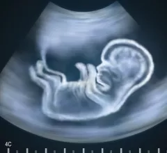
MRI should be a standard diagnostic tool for fetuses with known congenital heart disease

The modality can detect certain anomalies difficult to spot using only ultrasound, researchers reported in the Journal of the American College of Cardiology.

Around the web

The nuclear imaging isotope shortage of molybdenum-99 may be over now that the sidelined reactor is restarting. ASNC's president says PET and new SPECT technologies helped cardiac imaging labs better weather the storm.

CMS has more than doubled the CCTA payment rate from $175 to $357.13. The move, expected to have a significant impact on the utilization of cardiac CT, received immediate praise from imaging specialists.

The newly cleared offering, AutoChamber, was designed with opportunistic screening in mind. It can evaluate many different kinds of CT images, including those originally gathered to screen patients for lung cancer. 

Trimed Popup
Trimed Popup