Magnetic Resonance Imaging

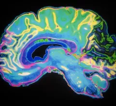
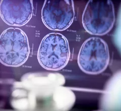
Magnetic resonance imaging (MRI) is the gold standard imaging modality for soft tissues. It produces detail cross-sectional images of soft tissue and bone anatomy, including muscles, tendons, ligaments, brain and organs, without the use of ionizing radiation. In addition to orthopedic imaging, MRI is also used for heart, brain and breast. MRI uses gadolinium contrast in many exams to highlight tissues and blood vessels, which enhances images and offers better diagnostic quality. It can also be used in conjunction with PET scans. How does MRI work? MR creates images by using powerful magnets to polarize hydrogen atoms in water (the body is made of of more than 80% water) so they face in one direction. A radiofrequency pulse is then used to ping these atoms, causing them to wobble, or resonate. The MRI coils detect this and computers can assemble images from the signals. Basic MRI scans will focus on the resonance of fat and water in two different sequences, which highlight and contrast different features in the anatomy.

coronavirus COVID-19 vaccine vaccination

Cardiac MRI scans offer new insight into COVID vaccine-related myocarditis

Vaccine-related myocarditis displayed less severe injury patterns than the heart inflammation caused by COVID infection, doctors explained in Radiology.

Thumbnail

These 3 MRI features can help junior radiologists identify placental attachment disorders

When using three specific MRI features to identify placental attachment abnormalities, junior radiologists' detection accuracy increased significantly, experts recently explained in EJR.

Thumbnail

Is PET/CT or multiparametric MRI best for detecting prostate cancer? New research offers guidance

A comparison of the detection rates for 68Ga-PSMA PET/CT and mpMRI in low, intermediate and high-risk prostate cancer was recently shared by experts in EJNMMI.

University awarded research grant to study Alzheimer's using specialized brain MRI

The $3.8 million grant spans five years and will focus on cerebrovascular abnormalities by using a specialized imaging technique, the university announced this week.

Wearable MRI helmet makes brain scans quicker, quieter and crisper

The helmet is made from magnetic metamaterials and can cut brain scan times in half, researchers at Boston University recently explained.

Thumbnail

'Highly significant' MRI findings link hyperthyroidism to structural brain abnormalities

“For decades, the patients in our group have testified that they don’t feel they’ve recovered, and we hope our study will provide further clues about what happens in the brain,” experts involved in the study said.

Thumbnail

Previous negative MRI experience significantly impacts patient anxiety during scans

“Beyond causing a negative patient experience, stress and anxiety also relate to unexpected patient-related events, which delay the clinical workflow and effect a significant amount of lost revenue," experts said in JACR.

MR-guided thermoseeds can destroy cancerous brain tissue

"Improving the precision of treatment delivery is arguably the greatest unmet need we have in contemporary medicine," experts explained in Advanced Science.

Around the web

The nuclear imaging isotope shortage of molybdenum-99 may be over now that the sidelined reactor is restarting. ASNC's president says PET and new SPECT technologies helped cardiac imaging labs better weather the storm.

CMS has more than doubled the CCTA payment rate from $175 to $357.13. The move, expected to have a significant impact on the utilization of cardiac CT, received immediate praise from imaging specialists.

The newly cleared offering, AutoChamber, was designed with opportunistic screening in mind. It can evaluate many different kinds of CT images, including those originally gathered to screen patients for lung cancer. 

Trimed Popup
Trimed Popup