Magnetic Resonance Imaging

Magnetic resonance imaging (MRI) is the gold standard imaging modality for soft tissues. It produces detail cross-sectional images of soft tissue and bone anatomy, including muscles, tendons, ligaments, brain and organs, without the use of ionizing radiation. In addition to orthopedic imaging, MRI is also used for heart, brain and breast. MRI uses gadolinium contrast in many exams to highlight tissues and blood vessels, which enhances images and offers better diagnostic quality. It can also be used in conjunction with PET scans. How does MRI work? MR creates images by using powerful magnets to polarize hydrogen atoms in water (the body is made of of more than 80% water) so they face in one direction. A radiofrequency pulse is then used to ping these atoms, causing them to wobble, or resonate. The MRI coils detect this and computers can assemble images from the signals. Basic MRI scans will focus on the resonance of fat and water in two different sequences, which highlight and contrast different features in the anatomy.

Philips unveils new radiation oncology portfolio for a more confident path to treatment at ASTRO 2018

New Big Bore RT and Ingenia Ambition/Elition MR-RT imaging systems and Pinnacle Evolution treatment planning software support improved patient care, accelerated time to treatment and enhanced patient satisfaction.

Thumbnail

Can diffusion-weighted imaging supplement dynamic contrast-enhanced breast MRI?

Diffusion-weighted imaging (DWI) could help differentiate between benign and malignant tumors and predict tumor recurrence in breast cancer patients, according to research published Oct. 4 in Academic Radiology. The authors noted that DWI may also serve as a beneficial supplement to contrast-enhanced breast MRI.

Thumbnail

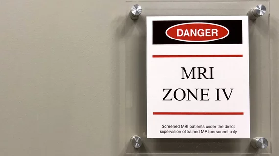
New York patient discharges gun in MRI room, charged with reckless endangerment

A Long Island, New York, man was arrested last month after a handgun he possessed fired and injured him while in an MRI room, according to a June 20 report from the LI Herald.

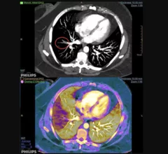
Example of spectral, or dual-energy CT, confirming a pulmonary embolism (PE). Image courtesy of Philips Healthcare

MRA vs CTA for evaluating pulmonary embolism: Does the chosen modality impact downstream imaging utilization?

MR angiography (MRA) is a relatively new alternative to CT angiography (CTA) for the evaluation of suspected pulmonary embolism (PE) and it even has one major advantage over CTA: it does not expose patients to ionizing radiation.

Example of pulmonary embolism (PE) detection on CT and confirmed using spectral CT. Image courtesy of Philips Healthcare

Researchers 'concerned' CT angiography topped MRA in follow-ups for pulmonary embolism

A recent study found whether a patient received MR angiography (MRA) instead of CT angiography (CTA) for a pulmonary embolism (PE) had little effect on the rate of follow-up chest CT or MRI one year later.

Image reconstruction algorithm, MRI-derived heart strain values can aid prognosis in amyloidosis patients

Recent research found strain parameters taken from a cine MRI-based deformable registration algorithm (DRA) can determine the severity of amyloid buildup in the heart and may provide prognostic information on patients with light-chain (AL) amyloidosis.

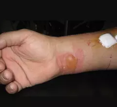
Example of a RF burn from an MRI scan where a sedated patient's identification bracelet was touching their skin during an exam. Image courtesy of RSNA. https://pubs.rsna.org/doi/10.1148/radiol.09090637

Battling Burns in MRI

While MRI is often considered one of the safer imaging modalities due to its lack of ionizing radiation, other dangers remain for patients, with thermal burns being a chief concern. 

Thumbnail

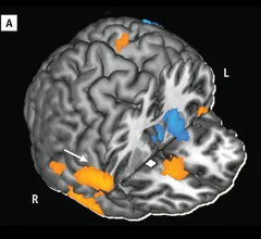
Imaging Addiction: Could PET & MR End Cocaine Abuse?

Cocaine addiction can ruin a person physically and financially, and with an estimated 1.4 million cocaine users in the U.S., thousands will become trapped by their habit. While previous research on the drug and its addictive potential were observational and subjective, imaging is reshaping how we see addiction—and how it will be treated.

Around the web

The nuclear imaging isotope shortage of molybdenum-99 may be over now that the sidelined reactor is restarting. ASNC's president says PET and new SPECT technologies helped cardiac imaging labs better weather the storm.

CMS has more than doubled the CCTA payment rate from $175 to $357.13. The move, expected to have a significant impact on the utilization of cardiac CT, received immediate praise from imaging specialists.

The newly cleared offering, AutoChamber, was designed with opportunistic screening in mind. It can evaluate many different kinds of CT images, including those originally gathered to screen patients for lung cancer. 

Trimed Popup
Trimed Popup