Magnetic Resonance Imaging

Magnetic resonance imaging (MRI) is the gold standard imaging modality for soft tissues. It produces detail cross-sectional images of soft tissue and bone anatomy, including muscles, tendons, ligaments, brain and organs, without the use of ionizing radiation. In addition to orthopedic imaging, MRI is also used for heart, brain and breast. MRI uses gadolinium contrast in many exams to highlight tissues and blood vessels, which enhances images and offers better diagnostic quality. It can also be used in conjunction with PET scans. How does MRI work? MR creates images by using powerful magnets to polarize hydrogen atoms in water (the body is made of of more than 80% water) so they face in one direction. A radiofrequency pulse is then used to ping these atoms, causing them to wobble, or resonate. The MRI coils detect this and computers can assemble images from the signals. Basic MRI scans will focus on the resonance of fat and water in two different sequences, which highlight and contrast different features in the anatomy.

Image reconstruction algorithm, MRI-derived heart strain values can aid prognosis in amyloidosis patients

Recent research found strain parameters taken from a cine MRI-based deformable registration algorithm (DRA) can determine the severity of amyloid buildup in the heart and may provide prognostic information on patients with light-chain (AL) amyloidosis.

Example of a RF burn from an MRI scan where a sedated patient's identification bracelet was touching their skin during an exam. Image courtesy of RSNA. https://pubs.rsna.org/doi/10.1148/radiol.09090637

Battling Burns in MRI

While MRI is often considered one of the safer imaging modalities due to its lack of ionizing radiation, other dangers remain for patients, with thermal burns being a chief concern. 

Thumbnail

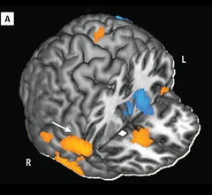
Imaging Addiction: Could PET & MR End Cocaine Abuse?

Cocaine addiction can ruin a person physically and financially, and with an estimated 1.4 million cocaine users in the U.S., thousands will become trapped by their habit. While previous research on the drug and its addictive potential were observational and subjective, imaging is reshaping how we see addiction—and how it will be treated.

Additional MR Techniques Improve Detection of Pulmonary Embolism

According a study published in the March issue of the journal Radiology, radiologists now have a comparable, non-ionizing option to CT for the detection of pulmonary embolism.

Radiology: Additional MR sequences improve pulmonary embolism detection

Adding two MRI sequences to a common MR pulmonary angiogram (MRPA) significantly improves detection of pulmonary embolism and could provide an alternative to CT angiography (CTA) for diagnosis, according to a study published in the April issue of Radiology.

Around the web

The new technology shows early potential to make a significant impact on imaging workflows and patient care. 

Richard Heller III, MD, RSNA board member and senior VP of policy at Radiology Partners, offers an overview of policies in Congress that are directly impacting imaging.
 

The two companies aim to improve patient access to high-quality MRI scans by combining their artificial intelligence capabilities.