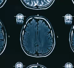
Magnetic Resonance Imaging

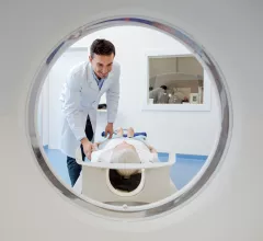
Magnetic resonance imaging (MRI) is the gold standard imaging modality for soft tissues. It produces detail cross-sectional images of soft tissue and bone anatomy, including muscles, tendons, ligaments, brain and organs, without the use of ionizing radiation. In addition to orthopedic imaging, MRI is also used for heart, brain and breast. MRI uses gadolinium contrast in many exams to highlight tissues and blood vessels, which enhances images and offers better diagnostic quality. It can also be used in conjunction with PET scans. How does MRI work? MR creates images by using powerful magnets to polarize hydrogen atoms in water (the body is made of of more than 80% water) so they face in one direction. A radiofrequency pulse is then used to ping these atoms, causing them to wobble, or resonate. The MRI coils detect this and computers can assemble images from the signals. Basic MRI scans will focus on the resonance of fat and water in two different sequences, which highlight and contrast different features in the anatomy.

Thumbnail

AI drops tracer dose required for PET/MRI by 50%, a potential ‘major’ boost for cancer care

The FDA-approved algorithm can ultimately minimize patients' risk of developing secondary cancers later in life, experts said in Radiology: Artificial Intelligence.

Thumbnail

AI triages breast screening MRIs without missing a single cancer

The system also sent nearly 90% of dense breast scans for radiologist review due to abnormal lesions.

Thumbnail

Radiology provider loses court battle with competitor over certificate of need for MRI

The North Carolina Court of Appeals acknowledged a loophole in the law but said its job is to interpret statutes as they are written, not speculate about consequences.

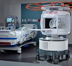
Portable MRI specialist Hyperfine announces plans to expand overseas

The move is part of a larger commercial expansion goal to enter four additional countries within the next six months.

Thumbnail

Pairing faster imaging sequence with deep learning cuts shoulder MRI scan time by 67%

Exams performed using the deep learning-based reconstruction tool also maintained high image quality, experts reported recently.

Thumbnail

MR imaging-guided SABR extends the lives of patients with inoperable pancreatic cancer

Miami Cancer Institute experts found the technique "significantly" prolonged overall survival, sharing their findings at the European Society for Radiotherapy and Oncology 2021 conference.

4D flow MRI has potential role for detecting complications in patients with aortic disease

One Northwestern Medicine researcher said the "landmark" findings take providers one step closer to using the tool for clinical decision making.

Thumbnail

Deep learning diagnoses disabling neurological condition on MRI in under 1 second

The platform—DystoniaNet—was 98.8% accurate at diagnosing the condition, and with further testing, may soon be in clinics, experts explained recently.

Around the web

Positron, a New York-based nuclear imaging company, will now provide Upbeat Cardiology Solutions with advanced PET/CT systems and services. 

The nuclear imaging isotope shortage of molybdenum-99 may be over now that the sidelined reactor is restarting. ASNC's president says PET and new SPECT technologies helped cardiac imaging labs better weather the storm.

CMS has more than doubled the CCTA payment rate from $175 to $357.13. The move, expected to have a significant impact on the utilization of cardiac CT, received immediate praise from imaging specialists.