Breast Imaging

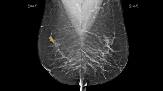
Breast imaging includes imaging modalities used for breast cancer screenings and planning therapy once cancer is detected. Mammography is the primary modality used. Mammogram technology is moving from 2D full-field digital mammography (FFDM) to breast tomosynthesis, or 3D mammography, which helps reduce false positive exams by allowing radiologists to look through the layers of tissue. Overlapping areas of dense breast tissue on 2D mammograms appear similar to cancers and 3D tomo helps determine if suspect areas are cancer or not. About 50% of women have dense breast tissue, which appears white on mammograms, the same as cancers, making diagnosis difficult. Radiologists use the Breast Imaging Reporting and Data System (BI-RADS) scoring system to define the density of breast tissue. Many states now require patients to be notified if they have dense breasts so they understand their mammograms might be suboptimal and they should use supplemental imaging that can see through the dense areas. This includes tomosythesis, breast ultrasound, automated breast ultrasound (ABUS), breast MRI, contrast enhanced mammography and nuclear imaging, including positron emission mammography (PEM).

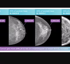
Illustration of the four types of breast tissue densities. The more dense, the harder it is for radiologists to detect cancers, which had led to about 40 states to now require notiofication of patients if they have dense breasts and the impact on their care, with possible miss-reads and that they may need supplemental imaging.

VIDEO: What is the impact of breast density notification laws?

Society of Breast Imaging (SBI) President John Lewin, MD, discusses how legislation concerning breast density notifications has impacted mammography.

Thumbnail

How breast cancer screening could increase lung cancer screening compliance in eligible women

Breast cancer screenings present an additional opportunity to identify more women who would also qualify for lung cancer screening, authors of a new paper in JAMA said.

black woman breast cancer pink ribbon

Disparities in breast cancer detection and care persist, despite a drop in mortality, new ACS report reveals

While Black women have lower incidence of breast cancer diagnosis, their mortality rates are 40% higher than those observed in white women.  

Thumbnail

Experts share positive news for women who have undergone multiple GBCA exposures during breast MRI

Authors of the new study stated that their findings are “reassuring” for both clinicians and patients.

Thumbnail

22% of eligible young women have not undergone a mammogram yet

Also, up to 26% of eligible women have not had a formal conversation with their physician regarding their breast cancer risks, despite current guidelines that suggest these discussions start sooner rather than later.

Thumbnail

Longitudinal changes in breast density could be linked to breast cancer risk

Researchers found that cancer risk in premenopausal women with fatty breasts at initial imaging nearly doubled if an increase in density was observed during their second and third mammograms.

Examples of breast imaging. What does breast imaging look like? What does breast cancer look like? What do radiologists look for on mammograms?

PHOTO GALLERY: What does breast cancer look like on mammography

This image gallery shows examples from various breast imaging modalities, including digital breast tomosynthesis, ultrasound and breast MRI, in addition to clinical presentations of breast cancer and other pathologies.

Experts describe MRI characteristics of rare breast cancer

The new paper describes the imaging characteristics of metaplastic carcinoma of the breast and correlates the findings with clinical and histopathological characteristics/diagnoses.

Around the web

Positron, a New York-based nuclear imaging company, will now provide Upbeat Cardiology Solutions with advanced PET/CT systems and services. 

The nuclear imaging isotope shortage of molybdenum-99 may be over now that the sidelined reactor is restarting. ASNC's president says PET and new SPECT technologies helped cardiac imaging labs better weather the storm.

CMS has more than doubled the CCTA payment rate from $175 to $357.13. The move, expected to have a significant impact on the utilization of cardiac CT, received immediate praise from imaging specialists.

Trimed Popup
Trimed Popup