Breast Imaging

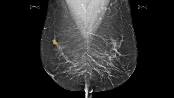
Breast imaging includes imaging modalities used for breast cancer screenings and planning therapy once cancer is detected. Mammography is the primary modality used. Mammogram technology is moving from 2D full-field digital mammography (FFDM) to breast tomosynthesis, or 3D mammography, which helps reduce false positive exams by allowing radiologists to look through the layers of tissue. Overlapping areas of dense breast tissue on 2D mammograms appear similar to cancers and 3D tomo helps determine if suspect areas are cancer or not. About 50% of women have dense breast tissue, which appears white on mammograms, the same as cancers, making diagnosis difficult. Radiologists use the Breast Imaging Reporting and Data System (BI-RADS) scoring system to define the density of breast tissue. Many states now require patients to be notified if they have dense breasts so they understand their mammograms might be suboptimal and they should use supplemental imaging that can see through the dense areas. This includes tomosythesis, breast ultrasound, automated breast ultrasound (ABUS), breast MRI, contrast enhanced mammography and nuclear imaging, including positron emission mammography (PEM).

Thumbnail

Many providers remain underinformed about increased cancer risks associated with breast density

JoAnn Pushkin, breast cancer survivor and executive director of DenseBreast-info.org, sounded the alarm on the lack of awareness surrounding breast density and its associated cancer risks during a presentation at this year’s annual RSNA meeting.

Thumbnail

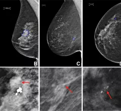
How breast cancer presents on imaging based on race: An updated analysis 25 years in the making

These appearances are important to recognize because, although cancer detection rates are similar among both races, mortality is approximately 40% higher among African American women in comparison to Non-Latina white women.

synthetic contrast-enhanced breast MRI

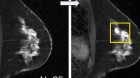
Are synthetic contrast-enhanced breast MRI images as good as the real thing?

Synthesizing contrast-enhanced imaging from pre-contrast inputs using deep learning can help overcome obstacles involved in the use of GBCAs.

womanconsultation.jpg

Personalized breast cancer risk assessment could reduce overdiagnosis

Personalized breast cancer screening programs could help women better understand their personal cancer risks and guide providers in recommending mammographic assessments based on these risks. 

Thumbnail

New data deepens debate over links between breast arterial calcification and CAD

The presence of breast arterial calcifications on mammography exams might not be as indicative of cardiovascular risk as previously thought, according to new data.

Thumbnail

Why some women tend to overestimate their breast cancer risks

For women with dense breasts, it can be particularly challenging to differentiate between actual and perceived breast cancer risks.  

AI identifies breast lesion subtypes, could prevent unnecessary biopsies

In a new paper in Radiology, experts explain how the use of artificial intelligence to identify lesion subtypes could benefit both clinics and patients. 

The FDA tracks mammography systems and the number of exams performed in the U.S. through its Mammography Quality Standards Act (MQSA) program. This is an example of the GE Senobright HD imaging system.

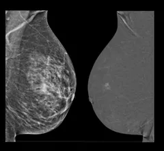
Is contrast-enhanced mammography suitable for women with breast implants?

A new paper highlights several studies supporting the use of screening mammography for women with breast implants, as well as additional considerations that need to be made for these patients.  

Around the web

Positron, a New York-based nuclear imaging company, will now provide Upbeat Cardiology Solutions with advanced PET/CT systems and services. 

The nuclear imaging isotope shortage of molybdenum-99 may be over now that the sidelined reactor is restarting. ASNC's president says PET and new SPECT technologies helped cardiac imaging labs better weather the storm.

CMS has more than doubled the CCTA payment rate from $175 to $357.13. The move, expected to have a significant impact on the utilization of cardiac CT, received immediate praise from imaging specialists.

Trimed Popup
Trimed Popup