Breast Imaging

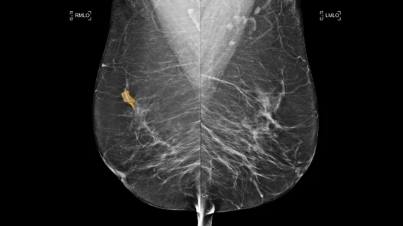
Breast imaging includes imaging modalities used for breast cancer screenings and planning therapy once cancer is detected. Mammography is the primary modality used. Mammogram technology is moving from 2D full-field digital mammography (FFDM) to breast tomosynthesis, or 3D mammography, which helps reduce false positive exams by allowing radiologists to look through the layers of tissue. Overlapping areas of dense breast tissue on 2D mammograms appear similar to cancers and 3D tomo helps determine if suspect areas are cancer or not. About 50% of women have dense breast tissue, which appears white on mammograms, the same as cancers, making diagnosis difficult. Radiologists use the Breast Imaging Reporting and Data System (BI-RADS) scoring system to define the density of breast tissue. Many states now require patients to be notified if they have dense breasts so they understand their mammograms might be suboptimal and they should use supplemental imaging that can see through the dense areas. This includes tomosythesis, breast ultrasound, automated breast ultrasound (ABUS), breast MRI, contrast enhanced mammography and nuclear imaging, including positron emission mammography (PEM).

Thumbnail

Should breast cancer screening initiation ages be reconsidered for women with a family history?

Suggesting that the standard 10-year age gap screening rule might not be as beneficial as previously assumed, researchers have indicated that women with relatives diagnosed at younger ages should consider different screening criteria.

Thumbnail

Prioritizing immediate reads on these two groups of women reduces recall rates for mammo

In a study cohort that included a total of 2,674,051 screening mammograms, these factors were found to result in the highest recall rates. 

Breast screening mammogram during the COVID pandemic. COVID significantly impacted breast imaging. Image courtesy of Novant Health

VIDEO: Impact of COVID on breast imaging

Connie Lehman, MD, PhD, chief of breast imaging, co-director of the Avon Comprehensive Breast Evaluation Center at the Massachusetts General Hospital, discusses the challenges of the COVID-19 pandemic on breast screenings, increased cancer rates and issues with the vaccines causing false positives on mammograms.

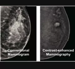
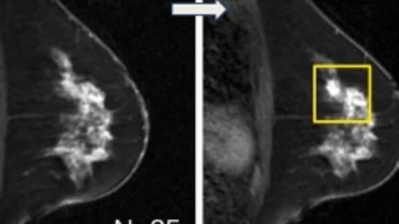
Contrast-enhanced mammography uses iodine contrast injected into a patient and mammography system to image contrast uptake or areas of increased vascular activity, which is typical of cancers. This can help image through dense breast tissue to find cancers that are otherwise masked by dense breast tissue.

VIDEO: Why contrast enhance mammography might be the ideal supplemental imaging

Connie Lehman, MD, PhD, chief of breast imaging, co-director of the Avon Comprehensive Breast Evaluation Center at the Massachusetts General Hospital, explains why she thinks contrast-enhanced mammography will likely become the go-to supplemental imaging modality for women with dense breasts.

MRI radiomics could change the future of breast cancer treatment

Radiomics methodologies could change how care plans are managed for patients with breast cancer by identifying those most likely to benefit from specific treatments.

Tattoo ink can mimic breast cancer on mammography exams

"As the prevalence of females with tattoos increases, tattoo pigment needs to be considered as a potential differential diagnosis of breast calcification,” experts involved in the research suggested.

An example of commercially available artificial intelligence (AI) automated grading of breast density on mammograms from the vendor Densitas..

VIDEO: Role of AI in breast imaging with radiomics, detection of breast density and lesions

Connie Lehman, MD, chief of breast imaging, co-director of the Avon Comprehensive Breast Evaluation Center at Massachusetts General Hospital, discusses how artificial intelligence (AI) is being implemented in breast imaging.

Example of a cancer that is difficult to see in dense breast tissue, but can be seen easier using 3D mammography digital breast tomosynthesis (DBT) breast imaging because the radiologist can go through the breast layer by layer if tissue..

VIDEO: The rapid adoption of 3D mammography and use of AI to address dense breasts

Stamatia Destounis, MD, a radiologist and managing partner at Elizabeth Wende Breast Care in Rochester, New York, chair of the American College of Radiology (ACR) Breast Commission, explains the rapid adoption of 3D mammogram digital breast tomosynthesis (DBT) technology.
 

Around the web

CMS has more than doubled the CCTA payment rate from $175 to $357.13. The move, expected to have a significant impact on the utilization of cardiac CT, received immediate praise from imaging specialists.

The newly cleared offering, AutoChamber, was designed with opportunistic screening in mind. It can evaluate many different kinds of CT images, including those originally gathered to screen patients for lung cancer. 

AI-enabled coronary plaque assessments deliver significant value, according to late-breaking data presented at TCT. These AI platforms have gained considerable momentum in recent months, receiving expanded Medicare coverage in addition to a new Category I CPT code.

Trimed Popup
Trimed Popup