Breast Imaging

Breast imaging includes imaging modalities used for breast cancer screenings and planning therapy once cancer is detected. Mammography is the primary modality used. Mammogram technology is moving from 2D full-field digital mammography (FFDM) to breast tomosynthesis, or 3D mammography, which helps reduce false positive exams by allowing radiologists to look through the layers of tissue. Overlapping areas of dense breast tissue on 2D mammograms appear similar to cancers and 3D tomo helps determine if suspect areas are cancer or not. About 50% of women have dense breast tissue, which appears white on mammograms, the same as cancers, making diagnosis difficult. Radiologists use the Breast Imaging Reporting and Data System (BI-RADS) scoring system to define the density of breast tissue. Many states now require patients to be notified if they have dense breasts so they understand their mammograms might be suboptimal and they should use supplemental imaging that can see through the dense areas. This includes tomosythesis, breast ultrasound, automated breast ultrasound (ABUS), breast MRI, contrast enhanced mammography and nuclear imaging, including positron emission mammography (PEM).

AI/radiologist combo improves breast cancer detection, decreases workloads

The new two-part AI system is based on a decision-referral approach and triages mammograms based on quantification of uncertainty.

Thumbnail

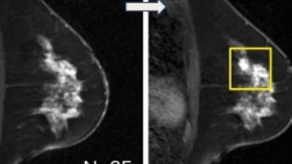
CEM vs breast MRI: Which is best for assessing treatment response?

A recent analysis of 51 patients revealed that contrast-enhanced mammography and MRI offered comparable assessments of lesion size, and both had similar specificity for pathologic complete response.

breast radiologist breast cancer mammography

Do race and ethnicity impact the accuracy of diagnostic mammograms?

There are many known disparities in breast imaging that have been found in previous studies, but insight into diagnostic mammography processes is still lacking. 

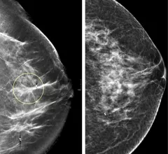
A comparison of standard 2D mammography (right) and digital breast tomosynthesis (DBT), or 3D mammography (left). The DBT creates a data set of 1 mm slices that the radiologist can look through to see more detail in suspect areas and determine if it dense breast tissue is masking a tumor.

DBT is better for women with increased breast cancer risks

For the research, special attention was paid to women with dense breasts and other factors that raise their risk of a breast cancer diagnosis.

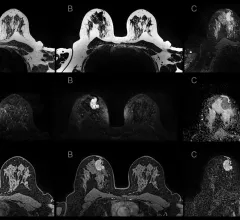
Example of various breast MRI protocol sequences that offer different types of soft tissue enhancement. Imaging performed on a Siemens Magnetom system. Breast MRI can help see through dense breast tissue to better detect cancers. #densebreasts #Breastdensity #BreastMRI

Contrast enhanced breast MRI superior to CEM, but accessibility remains an obstacle

In addition to CE-MRI's increased sensitivity for identifying breast cancers, the researchers also found the modality had superior negative likelihood ratios with higher pre-test probabilities for safely ruling out malignancy. 

Example of various breast MRI protocol sequences that offer different types of soft tissue enhancement. Imaging performed on a Siemens Magnetom system. Breast MRI can help see through dense breast tissue to better detect cancers. #densebreasts #Breastdensity #BreastMRI

MRI-based radiomics boosts triple-negative breast cancer detection

"The present study validated the feasibility of using MRI-based radiomics to identify a disease prone to missed diagnosis and misdiagnosis," experts involved in the study said.

A comparison of standard 2D mammography (right) and digital breast tomosynthesis (DBT), or 3D mammography (left). The DBT creates a data set of 1 mm slices that the radiologist can look through to see more detail in suspect areas and determine if it dense breast tissue is masking a tumor.

DBT not the superior modality for assessing BI-RADS 4 breast lesions

Assessing more than 11,000 patients with lesions designated as BI-RADS 4, radiologists using digital breast tomosynthesis found no significant diagnostic advantages over standard 2D mammography.

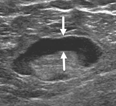
Breast ultrasound of a 62-year-old woman with benign reactive adenopathy after COVID-19 vaccination. Image from Academic Radiology.

Reactive lymphadenopathy slower to resolve after Moderna COVID vaccination

Imaging features were also examined as predictive time to resolution factors, with cortical thickness of the nodes being of particular interest.

Around the web

Positron, a New York-based nuclear imaging company, will now provide Upbeat Cardiology Solutions with advanced PET/CT systems and services. 

The nuclear imaging isotope shortage of molybdenum-99 may be over now that the sidelined reactor is restarting. ASNC's president says PET and new SPECT technologies helped cardiac imaging labs better weather the storm.

CMS has more than doubled the CCTA payment rate from $175 to $357.13. The move, expected to have a significant impact on the utilization of cardiac CT, received immediate praise from imaging specialists.

Trimed Popup
Trimed Popup