Breast Imaging

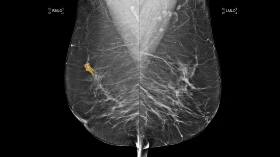
Breast imaging includes imaging modalities used for breast cancer screenings and planning therapy once cancer is detected. Mammography is the primary modality used. Mammogram technology is moving from 2D full-field digital mammography (FFDM) to breast tomosynthesis, or 3D mammography, which helps reduce false positive exams by allowing radiologists to look through the layers of tissue. Overlapping areas of dense breast tissue on 2D mammograms appear similar to cancers and 3D tomo helps determine if suspect areas are cancer or not. About 50% of women have dense breast tissue, which appears white on mammograms, the same as cancers, making diagnosis difficult. Radiologists use the Breast Imaging Reporting and Data System (BI-RADS) scoring system to define the density of breast tissue. Many states now require patients to be notified if they have dense breasts so they understand their mammograms might be suboptimal and they should use supplemental imaging that can see through the dense areas. This includes tomosythesis, breast ultrasound, automated breast ultrasound (ABUS), breast MRI, contrast enhanced mammography and nuclear imaging, including positron emission mammography (PEM).

Thumbnail

Lawmaker pushes to eliminate OOP costs for supplemental breast imaging

HB 575 would remove financial barriers for women who are recommended to undergo supplemental breast imaging, such as an MRI, CT or ultrasound, when the exams are deemed medically necessary.

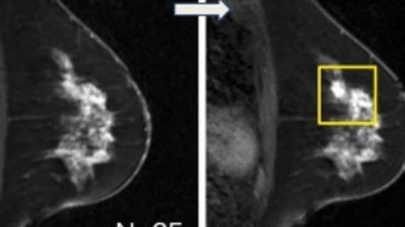
DCIS recurrence on mammogram

New data on DCIS surveillance imaging has experts concerned

A concerning number of women do not follow through with recommended surveillance imaging following treatment for breast cancer. 

 Winthrop P. Rockefeller Cancer Institute, University of Arkansas for Medical Sciences (USMS), is addressing health inequities in mammography using its Mammovan mobile breast imaging screening program. Gwendolyn Bryant-Smith, MD, explained how the program works. #RSNA #RSNA22

VIDEO: Bringing health equity to mammography and health screenings in Arkansas

Gwendolyn Bryant-Smith, MD, division chief of breast imaging and associate director for diversity, equity and inclusion at the Winthrop P. Rockefeller Cancer Institute, University of Arkansas, discussed how her center addressed health inequities in mammography.

Seno Medical's Imagio OA/US Breast Imaging System, optoacoustic ultrasound

Emerging optoacoustic ultrasound technology promises to reduce false-positives and unnecessary biopsies

Optoacoustic ultrasound can noninvasively provide valuable information on tumor behavior and better inform providers of whether a mass is benign or malignant. 

breast cancer screening using microwave breast imaging

Microwave breast imaging: A non-invasive, non-ionizing emerging technology for breast cancer screening

“This can have a major impact on the younger population, especially for women at high risk of developing breast cancer, like those with genetic mutations, where there is a necessity to start screenings at an early age,” experts explained in Academic Radiology.

womanconsultation.jpg

ACR recommendations result in 15-fold increase in breast MRI eligibility among cancer survivors

Prior to 2018, it was recommended that women at higher-than-average risk undergo supplemental breast MRI screening only if they had a personal history of breast cancer in addition to carrying a hereditary breast cancer gene mutation.

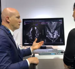
AI is still one of the key technologies on the floor many radiologists want to learn more about. of the AI. A product rep discussing breast automated detection AI in the crowded Lunit booth at RSNA 2022.

9 technology trends and takeaways from RSNA 2022

Here are some interesting new technologies and key trends from the vast expo floor at the Radiological Society of North America 2022 meeting.

Thumbnail

Follow-up recommendations after abnormal mammograms often ignored due to high deductibles

Although the Affordable Care Act eliminated OOP costs for screening mammograms under most insurance plans, that coverage does not carry over to the additional exams that are needed when abnormal findings are uncovered.

Around the web

Positron, a New York-based nuclear imaging company, will now provide Upbeat Cardiology Solutions with advanced PET/CT systems and services. 

The nuclear imaging isotope shortage of molybdenum-99 may be over now that the sidelined reactor is restarting. ASNC's president says PET and new SPECT technologies helped cardiac imaging labs better weather the storm.

CMS has more than doubled the CCTA payment rate from $175 to $357.13. The move, expected to have a significant impact on the utilization of cardiac CT, received immediate praise from imaging specialists.