Breast Imaging

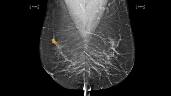
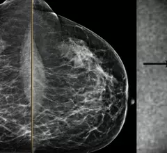
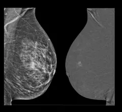
Breast imaging includes imaging modalities used for breast cancer screenings and planning therapy once cancer is detected. Mammography is the primary modality used. Mammogram technology is moving from 2D full-field digital mammography (FFDM) to breast tomosynthesis, or 3D mammography, which helps reduce false positive exams by allowing radiologists to look through the layers of tissue. Overlapping areas of dense breast tissue on 2D mammograms appear similar to cancers and 3D tomo helps determine if suspect areas are cancer or not. About 50% of women have dense breast tissue, which appears white on mammograms, the same as cancers, making diagnosis difficult. Radiologists use the Breast Imaging Reporting and Data System (BI-RADS) scoring system to define the density of breast tissue. Many states now require patients to be notified if they have dense breasts so they understand their mammograms might be suboptimal and they should use supplemental imaging that can see through the dense areas. This includes tomosythesis, breast ultrasound, automated breast ultrasound (ABUS), breast MRI, contrast enhanced mammography and nuclear imaging, including positron emission mammography (PEM).

mammography mammogram breast cancer

New data on false positive rates for DBT and DM screenings

Digital breast tomosynthesis exams produce fewer false positive results than standard two-dimensional mammography, though not substantially, experts shared recently in JAMA Open Network.

Thumbnail

The impact of state-level digital breast tomosynthesis coverage mandates

A total of 17 states have enacted legislation requiring private insurers to cover DBT exams, which has resulted in it being more widely recommended by radiologists. 

Thumbnail

States move breast cancer screening bills forward

The bills update the ages at which insurance carriers are required to cover screening mammograms and add language that includes tomosynthesis in the definition of mammographic screening.

Thumbnail

Breast density notifications leave many women in the dark, survey results suggest

Researchers are recommending a multi-modal approach to breast density education after survey results found that a "one size fits all" method fails many women.

Thumbnail

New AI tool accurately classifies breast density

The software was trained using more than 700 images and achieved a breast density classification accuracy of 89%, experts recently shared in Radiology: Artificial Intelligence.

Stacey Wolfson, MD, chief resident, and Beatriu Reig, MD, MPH, clinical assistant professor of radiology, Department of Radiology, NYU Grossman School of Medicine, explain the findings of a study they were the lead authors on published in Radiology. Their study looked at 1,200 women who were vaccinated and received breast imaging exams, and they found several cancers, so their conclusion is not to wait for breast imaging after receiving a COVID vaccine or booster.

VIDEO: Should women wait to get mammograms after COVID vaccination?

In an exclusive video, Stacey Wolfson, MD, and Beatriu Reig, MD, MPH, from the NYU Grossman School of Medicine, discuss the findings of their new analysis. 

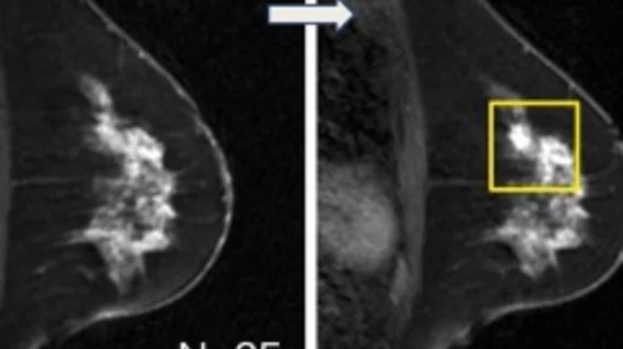
An example of a mammogram with some dense breast tissue that was deemed to not have cancer, and molecular breast imaging (MBI) study of the same women showing increased metabolic activity in the dense area, revealing a caner. Image from Mayo Clinic.

Mayo Clinic offers new guidance on supplemental screening of women with dense breasts

There are not yet consensus-based guidelines available for screening women with dense breast tissue, so researchers at Mayo Clinic recently developed a set of recommendations regarding supplemental screening. 

The FDA tracks mammography systems and the number of exams performed in the U.S. through its Mammography Quality Standards Act (MQSA) program. This is an example of the GE Senobright HD imaging system.

3D mammography approaching 50% of breast imaging systems in the U.S.

The latest U.S. Food and Drug Administration data on mammography systems installed nationwide shows DBT systems are rapidly replacing traditional 2D full field digital mammography units.

Around the web

Positron, a New York-based nuclear imaging company, will now provide Upbeat Cardiology Solutions with advanced PET/CT systems and services. 

The nuclear imaging isotope shortage of molybdenum-99 may be over now that the sidelined reactor is restarting. ASNC's president says PET and new SPECT technologies helped cardiac imaging labs better weather the storm.

CMS has more than doubled the CCTA payment rate from $175 to $357.13. The move, expected to have a significant impact on the utilization of cardiac CT, received immediate praise from imaging specialists.