Breast Imaging

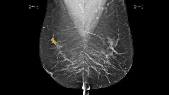
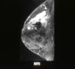
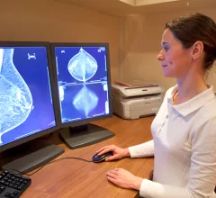
Breast imaging includes imaging modalities used for breast cancer screenings and planning therapy once cancer is detected. Mammography is the primary modality used. Mammogram technology is moving from 2D full-field digital mammography (FFDM) to breast tomosynthesis, or 3D mammography, which helps reduce false positive exams by allowing radiologists to look through the layers of tissue. Overlapping areas of dense breast tissue on 2D mammograms appear similar to cancers and 3D tomo helps determine if suspect areas are cancer or not. About 50% of women have dense breast tissue, which appears white on mammograms, the same as cancers, making diagnosis difficult. Radiologists use the Breast Imaging Reporting and Data System (BI-RADS) scoring system to define the density of breast tissue. Many states now require patients to be notified if they have dense breasts so they understand their mammograms might be suboptimal and they should use supplemental imaging that can see through the dense areas. This includes tomosythesis, breast ultrasound, automated breast ultrasound (ABUS), breast MRI, contrast enhanced mammography and nuclear imaging, including positron emission mammography (PEM).

Thumbnail

Machine learning uses MRI to predict lymphovascular invasion in breast cancer patients

These algorithms could fill in where postoperative biopsy sometimes falls short, experts explained in Academic Radiology.

Thumbnail

While promising, machine learning still misses 20% of cancers on breast MRIs, analysis shows

AI proved useful for detecting axillary lymph node metastases but isn’t yet ready for clinics, experts said recently.

Thumbnail

Providers are requesting fewer preoperative MRIs for breast cancer patients

Female surgeons, in particular, are abiding by evidence-based guidelines, but a more selective approach is still needed among doctors.

5 Years into the Cloud, John Muir Health Is Just Getting Started

Sponsored by Sectra

One 3D mammogram acquired via digital breast tomosynthesis adds about 500 MB of image data to a hospital’s storage system. That’s the average. On the high end, a single study can occupy as much as 3 GB of real estate on a finite-volume storage server.

Thumbnail

Ditching paper for e-worklist translates to ‘substantially’ shorter wait times for breast imaging patients

Brigham and Women's recently started using an electronic health record-embedded tool, resulting in many improvements for preoperative breast localization procedures.

Thumbnail

Breast radiologists’ opinions diverge on how to structure resident education, rotations

Imaging experts said they were "surprised" at the lack of consensus regarding how to manage first rotations during a 12-week program.

black woman breast cancer pink ribbon

Radiology advocates update breast cancer screening guidance to reflect higher risk for minority women

The American College of Radiology and Society of Breast Imaging noted women of color are 72% more likely to be diagnosed with the disease before they turn 50 compared to non-Hispanic white women.

Radiologists’ public service message underscores need to educate patients on COVID-19 vaccine side effects

Physicians called on imaging societies, clinicians and news media to spread awareness about vaccine-related swollen lymph nodes.

Around the web

Positron, a New York-based nuclear imaging company, will now provide Upbeat Cardiology Solutions with advanced PET/CT systems and services. 

The nuclear imaging isotope shortage of molybdenum-99 may be over now that the sidelined reactor is restarting. ASNC's president says PET and new SPECT technologies helped cardiac imaging labs better weather the storm.

CMS has more than doubled the CCTA payment rate from $175 to $357.13. The move, expected to have a significant impact on the utilization of cardiac CT, received immediate praise from imaging specialists.