Breast Imaging

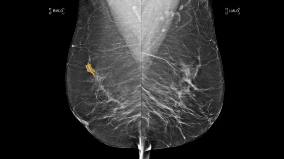
Breast imaging includes imaging modalities used for breast cancer screenings and planning therapy once cancer is detected. Mammography is the primary modality used. Mammogram technology is moving from 2D full-field digital mammography (FFDM) to breast tomosynthesis, or 3D mammography, which helps reduce false positive exams by allowing radiologists to look through the layers of tissue. Overlapping areas of dense breast tissue on 2D mammograms appear similar to cancers and 3D tomo helps determine if suspect areas are cancer or not. About 50% of women have dense breast tissue, which appears white on mammograms, the same as cancers, making diagnosis difficult. Radiologists use the Breast Imaging Reporting and Data System (BI-RADS) scoring system to define the density of breast tissue. Many states now require patients to be notified if they have dense breasts so they understand their mammograms might be suboptimal and they should use supplemental imaging that can see through the dense areas. This includes tomosythesis, breast ultrasound, automated breast ultrasound (ABUS), breast MRI, contrast enhanced mammography and nuclear imaging, including positron emission mammography (PEM).

Thumbnail

Fast breast MRI ready for clinical use following shorter read times, accurate cancer detection

Abbreviated MRI protocols not only shorten exam times but may significantly reduce costs and improve breast cancer screening overall.

COVID-19 globe coronavirus mask

All radiologists likely to encounter COVID-19 vaccine side effects in coming months, experts say

Massachusetts General Hospital rads outlined steps providers can take to limit unnecessary follow-up care in these situations, sharing their guidance in JACR.

mammography mammogram breast cancer

Here’s how radiologists should manage COVID-19 vaccine side effects spotted on breast MRI exams

Doctors have increasingly been seeing breast exams with swollen lymph nodes imitating cancer in patients who have received a vaccine, prompting Penn Medicine providers to offer up guidance.

Thumbnail

New suspicious findings on breast MRI performed during therapy are unlikely to be cancer

University of California, San Francisco, researchers found unusual lesions in 5.5% of cases and none were malignant.

Thumbnail

Breast MRI in cancer survivors results in more unneeded biopsies

Adding MRI to mammography screening for breast cancer detects more cancers, but results in more unnecessary biopsies, according to a June 4 study published in Radiology.

Thumbnail

Short-interval follow-up MRI helps ID early stage breast cancer

The researchers analyzed the frequency and cancer yield of ACR Breast Imaging Reporting and Data System (BI-RADS) 3 lesions in patients who received baseline and non-baseline screening MRIs.

Fujifilm launches three new software tools for its Aspire Cristalle mammography system

New features will be on display this spring at the National Consortium of Breast Centers conference and the Society of Breast Imaging Symposium

Thumbnail

Breast MRI may help personalize treatment for DCIS patients

Pairing breast MRI with a test that characterizes breast cancer genes can lead to a more personalized treatment approach for patients with ductal carcinoma in situ (DCIS), reported authors of a recent study published in JAMA Oncology.

Around the web

Positron, a New York-based nuclear imaging company, will now provide Upbeat Cardiology Solutions with advanced PET/CT systems and services. 

The nuclear imaging isotope shortage of molybdenum-99 may be over now that the sidelined reactor is restarting. ASNC's president says PET and new SPECT technologies helped cardiac imaging labs better weather the storm.

CMS has more than doubled the CCTA payment rate from $175 to $357.13. The move, expected to have a significant impact on the utilization of cardiac CT, received immediate praise from imaging specialists.