Breast Imaging

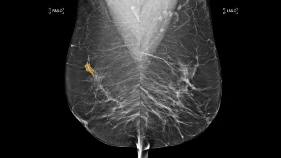
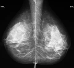
Breast imaging includes imaging modalities used for breast cancer screenings and planning therapy once cancer is detected. Mammography is the primary modality used. Mammogram technology is moving from 2D full-field digital mammography (FFDM) to breast tomosynthesis, or 3D mammography, which helps reduce false positive exams by allowing radiologists to look through the layers of tissue. Overlapping areas of dense breast tissue on 2D mammograms appear similar to cancers and 3D tomo helps determine if suspect areas are cancer or not. About 50% of women have dense breast tissue, which appears white on mammograms, the same as cancers, making diagnosis difficult. Radiologists use the Breast Imaging Reporting and Data System (BI-RADS) scoring system to define the density of breast tissue. Many states now require patients to be notified if they have dense breasts so they understand their mammograms might be suboptimal and they should use supplemental imaging that can see through the dense areas. This includes tomosythesis, breast ultrasound, automated breast ultrasound (ABUS), breast MRI, contrast enhanced mammography and nuclear imaging, including positron emission mammography (PEM).

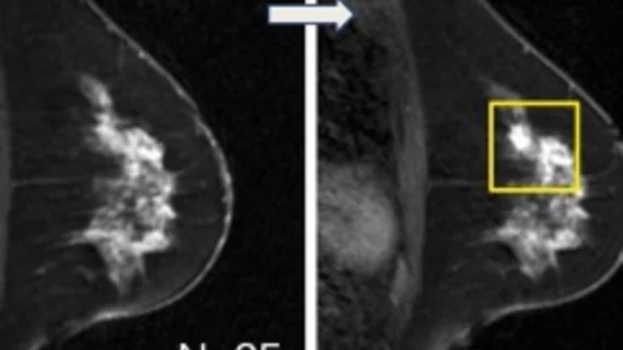
A new study suggests not delaying mammograms due to getting a COVID vaccine because cancers may go un detected. The RSNA study showed several examples of swollen lymph nodes that appeared to be from the vaccine, but turned out to be cancers.

Q&A: Should COVID vaccinated patients delay getting breast imaging — new study says no 

The lead authors of a new study, Stacey Wolfson, MD, and Beatriu Reig, MD, from the Department of Radiology, NYU Grossman School of Medicine, say women should not delay breast imaging if they recently received the COVID vaccine or a booster.

Thumbnail

Breast MRI screening cuts cancer mortality rates in half for women with lesser-known gene mutations

Women with ATM, CHEK2 and PALB2 gene mutations would benefit from beginning MRI screenings earlier than previously recommended, experts reported in JAMA Oncology.

Thumbnail

New research can help radiologists manage architectural distortion identified via DBT exams

After surgical excision, 10.2% of the architectural distortion cases with nonmalignant pathology at biopsy were upgraded to malignant, researchers reported in the American Journal of Roentgenology.

breast cancer mammography mammogram

Quality improvements reduce biopsy wait times for patients with suspicious breast MRIs

The initiative led to a 30% increase in the number of patients who required fewer visits before receiving a diagnosis, University of Michigan Health System researchers reported.

Thumbnail

Abbreviated MRI outperforms DBT for detecting secondary breast cancer

“Our study results suggest that AB-MRI could potentially be sufficient for post-treatment surveillance,” experts suggested Thursday in Academic Radiology.

Thumbnail

Machine learning model accurately predicts DCIS upstaging without invasive surgery

Understanding a patient's risk of developing invasive cancer without having to undergo surgery could help patients and providers choose more appropriate treatment plans.

breast ultrasound biopsy

Ultrasound outperforms four other modalities at assessing margins during breast surgery

Although ultrasound came out on top, achieving optimal operator performance could be taxing on resources, doctors cautioned.

Thumbnail

Deep learning rivals fellowship-trained radiologists at segmenting breast cancers on MRI

Researchers trained their platforms on more than 60,000 individual breast scans, significantly more than most architectures.

Around the web

Positron, a New York-based nuclear imaging company, will now provide Upbeat Cardiology Solutions with advanced PET/CT systems and services. 

The nuclear imaging isotope shortage of molybdenum-99 may be over now that the sidelined reactor is restarting. ASNC's president says PET and new SPECT technologies helped cardiac imaging labs better weather the storm.

CMS has more than doubled the CCTA payment rate from $175 to $357.13. The move, expected to have a significant impact on the utilization of cardiac CT, received immediate praise from imaging specialists.