Breast Imaging

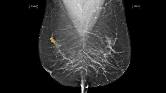
Breast imaging includes imaging modalities used for breast cancer screenings and planning therapy once cancer is detected. Mammography is the primary modality used. Mammogram technology is moving from 2D full-field digital mammography (FFDM) to breast tomosynthesis, or 3D mammography, which helps reduce false positive exams by allowing radiologists to look through the layers of tissue. Overlapping areas of dense breast tissue on 2D mammograms appear similar to cancers and 3D tomo helps determine if suspect areas are cancer or not. About 50% of women have dense breast tissue, which appears white on mammograms, the same as cancers, making diagnosis difficult. Radiologists use the Breast Imaging Reporting and Data System (BI-RADS) scoring system to define the density of breast tissue. Many states now require patients to be notified if they have dense breasts so they understand their mammograms might be suboptimal and they should use supplemental imaging that can see through the dense areas. This includes tomosythesis, breast ultrasound, automated breast ultrasound (ABUS), breast MRI, contrast enhanced mammography and nuclear imaging, including positron emission mammography (PEM).

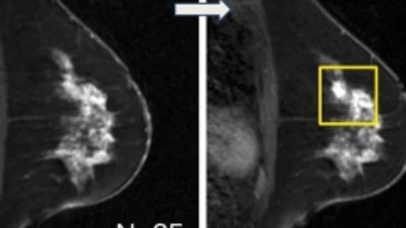
In preclinical trial, a new breast MRI contrast agent shows metastasis-prone cells at their earliest stage

Human trials are still a few years off, but a new contrast agent for early-detection breast MRI has come through biomedical imaging investigations in mice with flying colors. 

August 13, 2015

VolparaDensity Used to Guide Patient Selection in First Breast MRI Screening Trial for Women with Extremely Dense Breasts

The DENSE Trial, the first randomized trial investigating the additional value of MRI for screening women with dense breasts, is featured in the current issue of Radiology. 

July 8, 2015

Capitol briefing takes aim at USPSTF mammo guidelines

A congresswoman with a personal breast cancer story joined imaging experts for a Capitol Hill briefing that served both to illuminate the importance of mammography screening and also condemn recent U.S. Preventive Services Task Force mammography guidelines.

June 23, 2015

Volpara Solutions releases VolparaDensity 3.1 for use with tomosynthesis systems from multiple manufacturers at SBI

Volpara Solutions today announced the release of VolparaDensity version 3.1 here at the Society of Breast Imaging’s SBI/ACR Breast Imaging Symposium in Orlando, Fla., April 25-28, 2015 (SBI Booth #214).

April 28, 2015

Audits of breast MRI performance should separate screening from diagnostic

The cancer detection rate was significantly lower for breast MRI screenings than for diagnostic MRIs, according to a study published in the September issue of the Journal of the American College of Radiology.

September 4, 2014

Are breast MRIs and anxiety boosting rate of double mastectomies?

The rise in women getting double mastectomies after a breast cancer diagnosis could be linked to anxiety and breast MRI, as reported by NPR on May 21.

May 27, 2014
Example of a mammogram showing X-ray images of both the right and left breast and patches of dense breast tissue.

ACR, SBI launch screening mammo offense

A study published March 18 in Annals of Family Medicine that detailed long-term psychosocial harms of false-positive screening mammograms is compromised by methodological irregularities, underplays the harm of a breast cancer diagnosis and does not address existing strategies used to minimize anxiety, according to statements by the American College of Radiology (ACR), the Society of Breast Imaging (SBI) and breast imaging experts.

March 20, 2013
Example of a mammogram showing X-ray images of both the right and left breast and patches of dense breast tissue.

ACR & SBI: Norwegian mammo overdiagnosis analysis flawed

The American College of Radiology (ACR) and the Society of Breast Imaging (SBI) have responded to a study in the Annals of Internal Medicine which concluded that 15 to 25 percent of cancers in the Norwegian mammography screening program are overdiagnosed, saying those overdiagnosis rates are overestimated, according to a statement on ACRs website.

April 4, 2012

Around the web

Automated AI-generated measurements combined with annotated CT images can improve treatment planning and help referring physicians and patients better understand their disease, explained Sarah Jane Rinehart, MD, director of cardiac imaging with Charleston Area Medical Center.

Two advanced algorithms—one for CAC scores and another for segmenting cardiac chamber volumes—outperformed radiologists when assessing low-dose chest CT scans. 

"Gen AI can help tackle repetitive tasks and provide insights into massive datasets, saving valuable time," Thomas Kurian, CEO of Google Cloud, said Tuesday. 

Trimed Popup
Trimed Popup