Breast Imaging

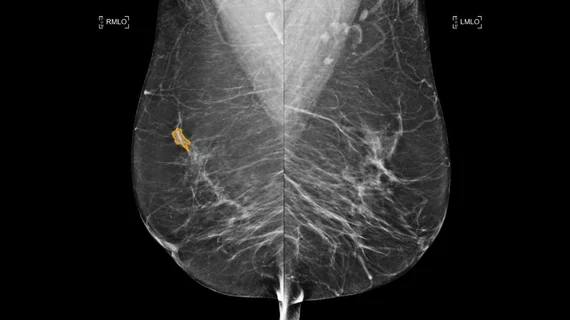
Breast imaging includes imaging modalities used for breast cancer screenings and planning therapy once cancer is detected. Mammography is the primary modality used. Mammogram technology is moving from 2D full-field digital mammography (FFDM) to breast tomosynthesis, or 3D mammography, which helps reduce false positive exams by allowing radiologists to look through the layers of tissue. Overlapping areas of dense breast tissue on 2D mammograms appear similar to cancers and 3D tomo helps determine if suspect areas are cancer or not. About 50% of women have dense breast tissue, which appears white on mammograms, the same as cancers, making diagnosis difficult. Radiologists use the Breast Imaging Reporting and Data System (BI-RADS) scoring system to define the density of breast tissue. Many states now require patients to be notified if they have dense breasts so they understand their mammograms might be suboptimal and they should use supplemental imaging that can see through the dense areas. This includes tomosythesis, breast ultrasound, automated breast ultrasound (ABUS), breast MRI, contrast enhanced mammography and nuclear imaging, including positron emission mammography (PEM).

Counseling increases mammo screening in low income populations

The most effective way to increase mammography screening among hard-to-reach, low-income, insured women is a stepwise intervention method or counseling program, according to a study published online June 29 in Cancer Epidemiology, Biomarkers & Prevention. The study found counseling increased screening compliance in the target population by 13.7 percent compared to the control group.

Molecular-based Breast Imaging: Catching Up to Breast MRI

Although breast MRI is an increasingly common complement to mammography because of its sensitivity in detecting suspicious lesions missed by mammography, two molecular imaging techniquespositron emission mammography (PEM) and breast specific gamma imaging (BSGI)are moving up the ranks due to greater physiological detail and spatial resolution both techniques offer.

iCAD nets 510(k) for interventional breast MRI app

The FDA has granted 510(k) clearance to iCAD for PrecisionPoint, its interventional planning solution for breast MRI that will be offered as an integrated module within the company's CADvue breast MRI image review software.

As CAD for Breast MRI & Mammography Gain Acceptance—Are Economic Issues the Final Barrier?

As more clinical studies tout the benefits of deploying computer-assisted detection (CAD) systems for breast MRI and mammography, the few lingering impediments to widespread adoption seem to be environmental. Low reimbursement rates, unfamiliarity with the technology and the troubled economy remain as potential barriers. However, most radiologists currently using the technology concur that both types of CAD systems add to the accuracy of detecting breast cancer, and therefore, allow for better patient care and almost immediate return on investment.

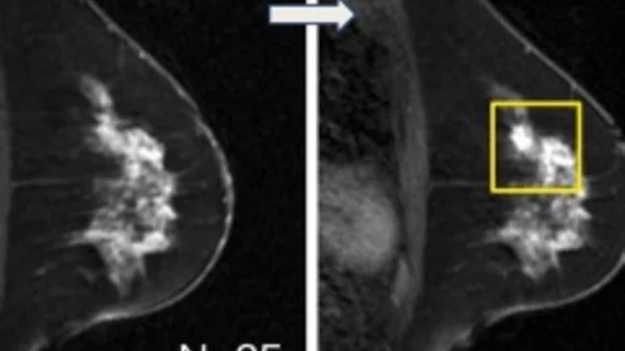
Breast MRI recommended for preoperative evaluation of newly diagnosed breast cancer

A study published in the American Journal of Surgery found that women who underwent breast MRI had a better chance of avoiding mastectomy and that the technology is a valid recommendation for preoperative evaluation of newly diagnosed breast cancer patients.

Two Aurora 1.5T breast MRI users gain MR accreditation

The Intersocietal Commission for the Accreditation of Magnetic Resonance Laboratories has accredited the Oklahoma City-based Breast MRI of Oklahoma and the Breast Center of Hattiesburg Clinic in Mississippi to use the 1.5Tesla breast MRI system from Aurora Imaging Technology.

Aurora nets 1.5T breast MRI install

Aurora Imaging Technology’s 1.5-Tesla dedicated breast MRI system isnow available to patients in northern Massachusetts and southern NewHampshire at Aurora Breast MRI of Beverly Hospital in Danvers, Mass.

Dedicated breast MRI leads growing adjunct breast imaging market

The market for adjunct breast imaging systems—covering breast coils,dedicated breast MRI systems and breast-specific gamma imaging systems—will grow at a compound annual growth rate of almost 28 percentover the next five years, according to a report from the MillenniumResearch Group.

Around the web

Positron, a New York-based nuclear imaging company, will now provide Upbeat Cardiology Solutions with advanced PET/CT systems and services. 

The nuclear imaging isotope shortage of molybdenum-99 may be over now that the sidelined reactor is restarting. ASNC's president says PET and new SPECT technologies helped cardiac imaging labs better weather the storm.

CMS has more than doubled the CCTA payment rate from $175 to $357.13. The move, expected to have a significant impact on the utilization of cardiac CT, received immediate praise from imaging specialists.