Breast Imaging

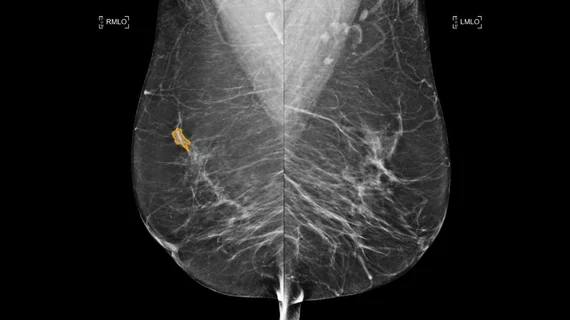
Breast imaging includes imaging modalities used for breast cancer screenings and planning therapy once cancer is detected. Mammography is the primary modality used. Mammogram technology is moving from 2D full-field digital mammography (FFDM) to breast tomosynthesis, or 3D mammography, which helps reduce false positive exams by allowing radiologists to look through the layers of tissue. Overlapping areas of dense breast tissue on 2D mammograms appear similar to cancers and 3D tomo helps determine if suspect areas are cancer or not. About 50% of women have dense breast tissue, which appears white on mammograms, the same as cancers, making diagnosis difficult. Radiologists use the Breast Imaging Reporting and Data System (BI-RADS) scoring system to define the density of breast tissue. Many states now require patients to be notified if they have dense breasts so they understand their mammograms might be suboptimal and they should use supplemental imaging that can see through the dense areas. This includes tomosythesis, breast ultrasound, automated breast ultrasound (ABUS), breast MRI, contrast enhanced mammography and nuclear imaging, including positron emission mammography (PEM).

Save time, catch cancer with abbreviated breast MRI protocols

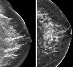
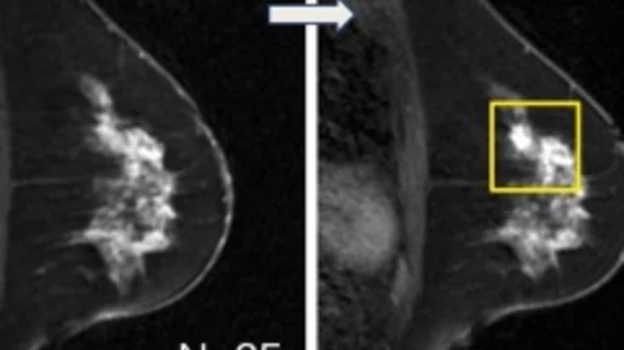
Abbreviated MRI protocols are just as effective as full diagnostic protocols at detecting cancers and also save valuable time and resources for both patients and facilities, according to results of a study published online in the Journal of the American College of Radiology.

Thumbnail

Study examines mismatch between breast MRI BI-RADS assessments, clinical recommendations

Breast MRI examinations assessed using BI-RADS guidelines don’t always coincide with expected clinical management recommendations, according to results of a study published online in the American Journal of Roentgenology.

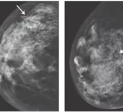
Architectural distortion seen in the breast of a 67-year-old woman who presented for screening mammography. Surgical pathology revealed invasive ductal adenocarcinoma. Image from AJR

When does worrisome architectural distortion signal malignancy on mammography?

Architectural distortion, the non-mass but potentially ominous clinical feature observed in many breast imaging procedures, is less likely to signal malignancy when it’s detected on screening mammography rather than diagnostic mammography or when it does not correlate with a subsequent targeted ultrasound exam.

Annual Mammography Starting at Age 40 Still Best Way to Saves Lives from Breast Cancer

The American College of Radiology (ACR), Society of Breast Imaging (SBI) and major medical organizations experienced in breast cancer care continue to recommend that women start getting annual mammograms at age 40.

Thumbnail

False-positives and fear

Research has once again shown that false-positive screening mammography results lead to increased anxiety for the women that receive them, and that these feelings can last for months. But how much should we fear the fear itself?

Thumbnail

In preclinical trial, a new breast MRI contrast agent shows metastasis-prone cells at their earliest stage

Human trials are still a few years off, but a new contrast agent for early-detection breast MRI has come through biomedical imaging investigations in mice with flying colors. 

VolparaDensity Used to Guide Patient Selection in First Breast MRI Screening Trial for Women with Extremely Dense Breasts

The DENSE Trial, the first randomized trial investigating the additional value of MRI for screening women with dense breasts, is featured in the current issue of Radiology. 

Thumbnail

Capitol briefing takes aim at USPSTF mammo guidelines

A congresswoman with a personal breast cancer story joined imaging experts for a Capitol Hill briefing that served both to illuminate the importance of mammography screening and also condemn recent U.S. Preventive Services Task Force mammography guidelines.

Around the web

CMS has more than doubled the CCTA payment rate from $175 to $357.13. The move, expected to have a significant impact on the utilization of cardiac CT, received immediate praise from imaging specialists.

The newly cleared offering, AutoChamber, was designed with opportunistic screening in mind. It can evaluate many different kinds of CT images, including those originally gathered to screen patients for lung cancer. 

AI-enabled coronary plaque assessments deliver significant value, according to late-breaking data presented at TCT. These AI platforms have gained considerable momentum in recent months, receiving expanded Medicare coverage in addition to a new Category I CPT code.

Trimed Popup
Trimed Popup