Breast Imaging

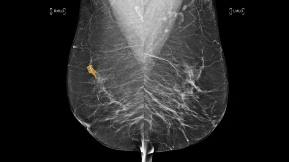
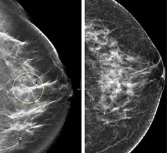
Breast imaging includes imaging modalities used for breast cancer screenings and planning therapy once cancer is detected. Mammography is the primary modality used. Mammogram technology is moving from 2D full-field digital mammography (FFDM) to breast tomosynthesis, or 3D mammography, which helps reduce false positive exams by allowing radiologists to look through the layers of tissue. Overlapping areas of dense breast tissue on 2D mammograms appear similar to cancers and 3D tomo helps determine if suspect areas are cancer or not. About 50% of women have dense breast tissue, which appears white on mammograms, the same as cancers, making diagnosis difficult. Radiologists use the Breast Imaging Reporting and Data System (BI-RADS) scoring system to define the density of breast tissue. Many states now require patients to be notified if they have dense breasts so they understand their mammograms might be suboptimal and they should use supplemental imaging that can see through the dense areas. This includes tomosythesis, breast ultrasound, automated breast ultrasound (ABUS), breast MRI, contrast enhanced mammography and nuclear imaging, including positron emission mammography (PEM).

AJR: Breast MRI protocol opens door to lymph node assessment

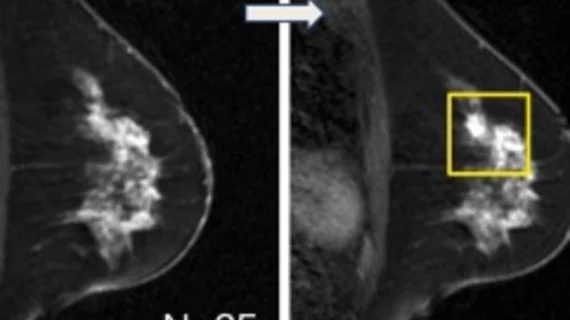
Combined local and locoregional staging via whole-body MRI is clinically feasible and can accurately evaluate axillary lymph nodes, providing data that may help physicians optimize management of breast cancer patients, according to a study published in the May issue of American Journal of Roentgenology.

Comparison of a 2D digital mammogram and breast tomosynthesis 3D mammography from UCSF.

ACR & SBI: Mammo not dangerous to thyroid

In response to an erroneous media report, the American College of Radiology (ACR) and the Society of Breast Imaging (SBI) released a joint statement urging patients to disregard trumped up risks of thyroid cancer due to mammography-induced radiation.

Radiology: False-positives drop w/ routine breast MRI screening

Women at high risk for breast cancer undergoing their first MRI screening have higher false-positive rates and require significantly more follow-up, showing that MRIs notoriously low specificity improves as women accumulate prior MR screening histories, a study published Feb. 1 in Radiology discovered.

RSNA: Penrad acquires Clarios Compass breast MRI software

PenRad Technologies (Booth 4600) has purchased Clario Medical Imaging's Compass breast MRI visualization and analysis software, which PenRad will launch at the annual meeting of the Radiological Society of North America (RSNA), Nov. 28 to Dec. 3, in Chicago.

Clinical Breast MRI

Clinical Breast MRI Sunday, Nov. 28, 2:00 PM - 3:30 PM

JACR: Community hospitals perform breast MRI well

A majority of community hospitals comply with two common sets of guidelines for high-quality breast MRI acquisition, despite the absence of uniform standards for breast MRI in the U.S., according to a survey of community hospitals published in the November edition of the Journal of the American College of Radiology (JACR).

AJR: Breast MRI screening beneficial for some with previous cancer

Breast MRI screening of women with only a personal, not familial, history of breast cancer was clinically valuable in finding malignancies for 12 percent of all cases, with a reasonable biopsy rate of 39 percent, found a study published in the August issue of the American Journal of Roentgenology.

Breast MRI: Looking Beyond High-risk Screening

Three years ago, the American Cancer Society (ACS) updated breast cancer screening guidelines for high-risk and dense-breasted, pre-menopausal women, recommending that this subset of patients undergo breast MRI in addition to annual screening mammograms.

Around the web

Positron, a New York-based nuclear imaging company, will now provide Upbeat Cardiology Solutions with advanced PET/CT systems and services. 

The nuclear imaging isotope shortage of molybdenum-99 may be over now that the sidelined reactor is restarting. ASNC's president says PET and new SPECT technologies helped cardiac imaging labs better weather the storm.

CMS has more than doubled the CCTA payment rate from $175 to $357.13. The move, expected to have a significant impact on the utilization of cardiac CT, received immediate praise from imaging specialists.