Breast Imaging

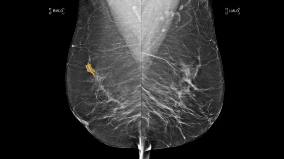
Breast imaging includes imaging modalities used for breast cancer screenings and planning therapy once cancer is detected. Mammography is the primary modality used. Mammogram technology is moving from 2D full-field digital mammography (FFDM) to breast tomosynthesis, or 3D mammography, which helps reduce false positive exams by allowing radiologists to look through the layers of tissue. Overlapping areas of dense breast tissue on 2D mammograms appear similar to cancers and 3D tomo helps determine if suspect areas are cancer or not. About 50% of women have dense breast tissue, which appears white on mammograms, the same as cancers, making diagnosis difficult. Radiologists use the Breast Imaging Reporting and Data System (BI-RADS) scoring system to define the density of breast tissue. Many states now require patients to be notified if they have dense breasts so they understand their mammograms might be suboptimal and they should use supplemental imaging that can see through the dense areas. This includes tomosythesis, breast ultrasound, automated breast ultrasound (ABUS), breast MRI, contrast enhanced mammography and nuclear imaging, including positron emission mammography (PEM).

Optimizing Breast MRI Reading

From the latest software to meticulous scheduling processes, breastimaging facilities are optimizing their efficiency when it comes toperforming and interpreting breast imaging studies.

Texas imaging facility buys Aurora breast MRI system

Aurora Imaging Technology’s 1.5Tesla Dedicated Breast MRI System is nowavailable at Wellness Lubbock, a women’s imaging facility in Lubbock,Texas.

Florida women?s imaging center to offer Aurora Breast MRI

Aurora Imaging Technology's 1.5Tesla dedicated breast MRI System is now available to Floridians at the Bay Radiology Women's Imaging Center in Panama City, Fla.

Texas cancer center installs Aurora breast MRI

The Comprehensive Breast Center of The Don and Sybil Harrington CancerCenter in Amarillo, Texas, has installed a 1.5Tesla dedicated BreastMRI System from Aurora Imaging Technology.

Aurora wins breast MRI install in Indiana

Breast MRI North in the Breast Diagnostic Center of Community HospitalNorth, a joint venture with Irvington Radiologists and CommunityHospital North, in Indianapolis, has installed the Aurora 1.5 Tesladedicated breast MRI from Aurora Imaging Technology.

Breast MRI before breast-conservation treatment does not improve outcome

The use of a breast MRI study at the time of initial diagnosis andevaluation was not associated with an improvement in outcome afterbreast-conservation treatment with radiation, according to astudy in the Jan. 20 issue of the Journal of Clinical Oncology.

Confirmas CADstream helpful for evaluating kinetics in breast MRI

Dec. 3—Confirma's CADstream system's patented "worst curve" algorithm for analyzing kinetics may assist in determining malignancy, according to a study presented by Lilian Wang, MD, of the radiology resident program at the Seattle Cancer Care Alliance and University of Washington in Seattle, at the 93rd scientific assembly and annual meeting of the Radiological Society of North America (RSNA) in Chicago last week.

Confirma, Penrad collaborating to on 1st breast MRI-mammography integration

CHICAGO, Nov. 27—Confirma, an application-specific computer-assisteddetection (CAD) company for MRI, and Penrad Technologies, a mammographyreporting company, are integrating between Confirma’s CADstream forbreast MRI and Penrad’s mammography information system, which is beingshowcased this week at the 93rd annual meeting of the RadiologicalSociety of North America (RSNA).

Around the web

Positron, a New York-based nuclear imaging company, will now provide Upbeat Cardiology Solutions with advanced PET/CT systems and services. 

The nuclear imaging isotope shortage of molybdenum-99 may be over now that the sidelined reactor is restarting. ASNC's president says PET and new SPECT technologies helped cardiac imaging labs better weather the storm.

CMS has more than doubled the CCTA payment rate from $175 to $357.13. The move, expected to have a significant impact on the utilization of cardiac CT, received immediate praise from imaging specialists.