Breast Imaging

Breast imaging includes imaging modalities used for breast cancer screenings and planning therapy once cancer is detected. Mammography is the primary modality used. Mammogram technology is moving from 2D full-field digital mammography (FFDM) to breast tomosynthesis, or 3D mammography, which helps reduce false positive exams by allowing radiologists to look through the layers of tissue. Overlapping areas of dense breast tissue on 2D mammograms appear similar to cancers and 3D tomo helps determine if suspect areas are cancer or not. About 50% of women have dense breast tissue, which appears white on mammograms, the same as cancers, making diagnosis difficult. Radiologists use the Breast Imaging Reporting and Data System (BI-RADS) scoring system to define the density of breast tissue. Many states now require patients to be notified if they have dense breasts so they understand their mammograms might be suboptimal and they should use supplemental imaging that can see through the dense areas. This includes tomosythesis, breast ultrasound, automated breast ultrasound (ABUS), breast MRI, contrast enhanced mammography and nuclear imaging, including positron emission mammography (PEM).

Volpara Solutions releases VolparaDensity 3.1 for use with tomosynthesis systems from multiple manufacturers at SBI

Volpara Solutions today announced the release of VolparaDensity version 3.1 here at the Society of Breast Imaging’s SBI/ACR Breast Imaging Symposium in Orlando, Fla., April 25-28, 2015 (SBI Booth #214).

Audits of breast MRI performance should separate screening from diagnostic

The cancer detection rate was significantly lower for breast MRI screenings than for diagnostic MRIs, according to a study published in the September issue of the Journal of the American College of Radiology.

Thumbnail

Are breast MRIs and anxiety boosting rate of double mastectomies?

The rise in women getting double mastectomies after a breast cancer diagnosis could be linked to anxiety and breast MRI, as reported by NPR on May 21.

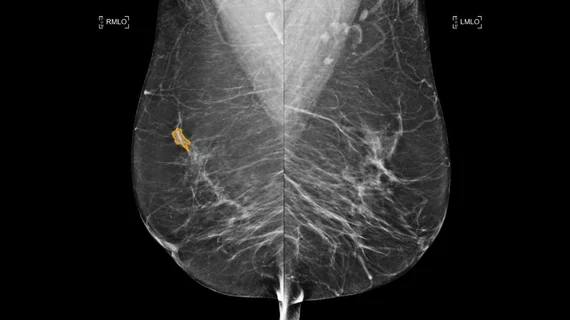
Example of a mammogram showing X-ray images of both the right and left breast and patches of dense breast tissue.

ACR, SBI launch screening mammo offense

A study published March 18 in Annals of Family Medicine that detailed long-term psychosocial harms of false-positive screening mammograms is compromised by methodological irregularities, underplays the harm of a breast cancer diagnosis and does not address existing strategies used to minimize anxiety, according to statements by the American College of Radiology (ACR), the Society of Breast Imaging (SBI) and breast imaging experts.

Example of a mammogram showing X-ray images of both the right and left breast and patches of dense breast tissue.

ACR & SBI: Norwegian mammo overdiagnosis analysis flawed

The American College of Radiology (ACR) and the Society of Breast Imaging (SBI) have responded to a study in the Annals of Internal Medicine which concluded that 15 to 25 percent of cancers in the Norwegian mammography screening program are overdiagnosed, saying those overdiagnosis rates are overestimated, according to a statement on ACRs website.

Comparison of a 2D digital mammogram and breast tomosynthesis 3D mammography from UCSF.

ACR & SBI: Mammo not dangerous to thyroid

In response to an erroneous media report, the American College of Radiology (ACR) and the Society of Breast Imaging (SBI) released a joint statement urging patients to disregard trumped up risks of thyroid cancer due to mammography-induced radiation.

Counseling increases mammo screening in low income populations

The most effective way to increase mammography screening among hard-to-reach, low-income, insured women is a stepwise intervention method or counseling program, according to a study published online June 29 in Cancer Epidemiology, Biomarkers & Prevention. The study found counseling increased screening compliance in the target population by 13.7 percent compared to the control group.

Around the web

CMS has more than doubled the CCTA payment rate from $175 to $357.13. The move, expected to have a significant impact on the utilization of cardiac CT, received immediate praise from imaging specialists.

The newly cleared offering, AutoChamber, was designed with opportunistic screening in mind. It can evaluate many different kinds of CT images, including those originally gathered to screen patients for lung cancer. 

AI-enabled coronary plaque assessments deliver significant value, according to late-breaking data presented at TCT. These AI platforms have gained considerable momentum in recent months, receiving expanded Medicare coverage in addition to a new Category I CPT code.

Trimed Popup
Trimed Popup