Breast Imaging

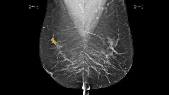
Breast imaging includes imaging modalities used for breast cancer screenings and planning therapy once cancer is detected. Mammography is the primary modality used. Mammogram technology is moving from 2D full-field digital mammography (FFDM) to breast tomosynthesis, or 3D mammography, which helps reduce false positive exams by allowing radiologists to look through the layers of tissue. Overlapping areas of dense breast tissue on 2D mammograms appear similar to cancers and 3D tomo helps determine if suspect areas are cancer or not. About 50% of women have dense breast tissue, which appears white on mammograms, the same as cancers, making diagnosis difficult. Radiologists use the Breast Imaging Reporting and Data System (BI-RADS) scoring system to define the density of breast tissue. Many states now require patients to be notified if they have dense breasts so they understand their mammograms might be suboptimal and they should use supplemental imaging that can see through the dense areas. This includes tomosythesis, breast ultrasound, automated breast ultrasound (ABUS), breast MRI, contrast enhanced mammography and nuclear imaging, including positron emission mammography (PEM).

Thumbnail

Another challenge to routine preop breast MRI

Those advocating against the routine use of preoperative breast MRI—an issue that has been in debate for years—received some more support in the literature this week, as a meta-analysis found no evidence that preoperative MRI for staging the cancerous breast reduces local or distant recurrence.

Thumbnail

Growth of breast MRI runs afoul of guidelines

Breast MRI use grew quickly in the early 2000s before stabilizing in recent years, but questions remain about whether American Cancer Society criteria for use are being followed, according to a study published online Nov. 18 in JAMA Internal Medicine.

Pre-op breast MRI linked to increasing rates of invasive surgery

Increasing use of preoperative breast MRI has been associated with significant increases in bilateral cancer diagnoses and subsequent invasive surgeries with questionable long-term benefits, according to a study published online Aug. 14 in Breast Cancer Research and Treatment.

Thumbnail

Mixed bag: Breast MRI CAD boosts sensitivity but not accuracy

A multicenter reader study designed to examine the effect of CAD on the diagnostic accuracy of breast MRI indicated improved sensitivity and comparable accuracy among novice and expert readers reviewing images without and with CAD, according to a study published in the June issue of American Journal of Roentgenology. CAD software did not increase interpretation time.

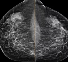
Example of a mammogram showing X-ray images of both the right and left breast and patches of dense breast tissue.

ACR, SBI launch screening mammo offense

A study published March 18 in Annals of Family Medicine that detailed long-term psychosocial harms of false-positive screening mammograms is compromised by methodological irregularities, underplays the harm of a breast cancer diagnosis and does not address existing strategies used to minimize anxiety, according to statements by the American College of Radiology (ACR), the Society of Breast Imaging (SBI) and breast imaging experts.

Pre-operative Breast MRI: Pros and Cons

Tuesday, Nov. 27, 7:15-8:15 a.m.| E451B | This session identifies areas of controversy surrounding pre-operative breast MRI and evidence both for and against its use. The presentations compare advantages and disadvantages of pre-operative MRI for women with newly diagnosed breast cancer.

Example of a mammogram showing X-ray images of both the right and left breast and patches of dense breast tissue.

ACR & SBI: Norwegian mammo overdiagnosis analysis flawed

The American College of Radiology (ACR) and the Society of Breast Imaging (SBI) have responded to a study in the Annals of Internal Medicine which concluded that 15 to 25 percent of cancers in the Norwegian mammography screening program are overdiagnosed, saying those overdiagnosis rates are overestimated, according to a statement on ACRs website.

AACR: Breast MRI screening cuts diagnostic costs for high-risk women

The use of breast MRI screenings among targeted, high-risk, underserved women significantly decreased diagnostic cost and increased patient follow-up compliance rates compared to using general-risk mammography screenings, according to a study presented at the Fourth American Association for Cancer Research (AACR) Conference on The Science of Cancer Health Disparities, held in Washington, D.C., from Sept. 18-21.

Around the web

Positron, a New York-based nuclear imaging company, will now provide Upbeat Cardiology Solutions with advanced PET/CT systems and services. 

The nuclear imaging isotope shortage of molybdenum-99 may be over now that the sidelined reactor is restarting. ASNC's president says PET and new SPECT technologies helped cardiac imaging labs better weather the storm.

CMS has more than doubled the CCTA payment rate from $175 to $357.13. The move, expected to have a significant impact on the utilization of cardiac CT, received immediate praise from imaging specialists.