Magnetic Resonance Imaging

Magnetic resonance imaging (MRI) is the gold standard imaging modality for soft tissues. It produces detail cross-sectional images of soft tissue and bone anatomy, including muscles, tendons, ligaments, brain and organs, without the use of ionizing radiation. In addition to orthopedic imaging, MRI is also used for heart, brain and breast. MRI uses gadolinium contrast in many exams to highlight tissues and blood vessels, which enhances images and offers better diagnostic quality. It can also be used in conjunction with PET scans. How does MRI work? MR creates images by using powerful magnets to polarize hydrogen atoms in water (the body is made of of more than 80% water) so they face in one direction. A radiofrequency pulse is then used to ping these atoms, causing them to wobble, or resonate. The MRI coils detect this and computers can assemble images from the signals. Basic MRI scans will focus on the resonance of fat and water in two different sequences, which highlight and contrast different features in the anatomy.

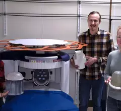
NIST researchers improve portable MRI

Improving low-field MRI scanners with quantitative data

Researchers at the National Institute of Standards and Technology (NIST) are working on developing measurements that will provide better insight into soft tissue properties visualized on portable MRI images. 

Thumbnail

New discovery paves the way for copper's entrance into MRI contrast agents

The new discovery contradicts the notion that copper is not suitable for use in contrast media.

Thumbnail

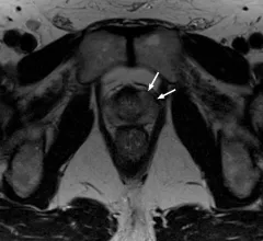
Preop prostate MRI findings predict cancer recurrence at rates similar to pathology, analysis shows

Given the similarities in accuracy between pathology results and preoperative imaging, MRI could have a greater role in risk assessments in the future, experts suggested in AJR.

Lumbar MRI exam duration cut in half using deep learning-based reconstruction algorithm

And the improved scan times did not come at the expense of image quality but, instead, offered improved signal-to-noise ratio, according to a new study in Skeletal Radiology.

Thumbnail

Poverty can change the wiring of a child's brain, MRI study suggests

New research out of the Washington University School of Medicine in St. Louis is offering new insight into how childhood struggles can affect brain development and connectivity. 

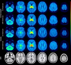
molecular imaging study on brain connections in obese individuals

Obesity linked to altered brain connectivity on PET/MRI exams

New research, presented at SNMMI 2023, details how the brains of obese individuals differ from those who maintain a healthy weight.  

MRA for pulmonary embolus

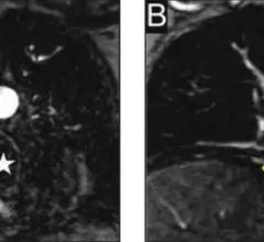
MR angiography a suitable alternative to CT when ruling out pulmonary embolus

The modality switch became especially important during the iodinated contrast shortage of 2022 when clinics were forced to deploy mitigation tactics as a means of preserving their contrast supply. 

New MRI technique offers detailed view of COVID's impact on the brain

The new technique mixes MRI signals at different gradient pulse strengths and timings, offering better visualization of differences in the way water molecules move through tissues.  

Around the web

Positron, a New York-based nuclear imaging company, will now provide Upbeat Cardiology Solutions with advanced PET/CT systems and services. 

The nuclear imaging isotope shortage of molybdenum-99 may be over now that the sidelined reactor is restarting. ASNC's president says PET and new SPECT technologies helped cardiac imaging labs better weather the storm.

CMS has more than doubled the CCTA payment rate from $175 to $357.13. The move, expected to have a significant impact on the utilization of cardiac CT, received immediate praise from imaging specialists.