Magnetic Resonance Imaging

Magnetic resonance imaging (MRI) is the gold standard imaging modality for soft tissues. It produces detail cross-sectional images of soft tissue and bone anatomy, including muscles, tendons, ligaments, brain and organs, without the use of ionizing radiation. In addition to orthopedic imaging, MRI is also used for heart, brain and breast. MRI uses gadolinium contrast in many exams to highlight tissues and blood vessels, which enhances images and offers better diagnostic quality. It can also be used in conjunction with PET scans. How does MRI work? MR creates images by using powerful magnets to polarize hydrogen atoms in water (the body is made of of more than 80% water) so they face in one direction. A radiofrequency pulse is then used to ping these atoms, causing them to wobble, or resonate. The MRI coils detect this and computers can assemble images from the signals. Basic MRI scans will focus on the resonance of fat and water in two different sequences, which highlight and contrast different features in the anatomy.

Imaging required to monitor effects of new Alzheimer's drug could inhibit its uptake

Leqembi can cause amyloid-related imaging abnormalities (ARIA), which must be assessed on imaging.

MRI-themed escape room University of Oxford

University creates MRI-themed escape room

The unique and immersive experience begins with visitors being met with an urgent warning that an AI "evil brain” has begun to operate independently and with hostility, threatening to put the laboratory into lockdown.

FDA issues adverse event report on MRI accident as investigations into its cause continue

The FDA's description of the event aligns with prior reports speculating that the accident involved a staff member transporting a patient to an MRI suite on a hospital bed, which was allegedly sucked into the MRI bore.

Thumbnail

In rare cases, maternal COVID infections can lead to serious brain injuries in neonates

Children included in the assessment displayed several concerning neurological MRI findings shortly after their birth, including acquired microcephaly, severe parenchymal atrophy and cystic encephalomalacia. 

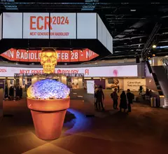
The European Society of radiology European Congress of Radiology (ECR) 2023 meeting. Image courtesy of ECR

Key trends in radiology at the European Congress of Radiology 2023 meeting

Bhvita Jani, research manager at the healthcare market analysis firm Signify Research, shares noteworthy happenings from the ECR expo floor.

Thumbnail

Experts identify sex-specific MRI brain signatures associated with obesity

Researchers at the University of California Los Angeles combined multimodal brain MRIs with clinical and personal data from 183 men and women to identify sex-specific brain signatures that could be indicative of what causes a person to develop obesity.

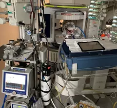
portable MRI in pediatric ECMO setting

Portable MRI achieves 'immense success' in study of critically ill children

Results from the world’s first study on the use of portable MRI equipment during pediatric ECMO therapy were published April 4 in Critical Care.

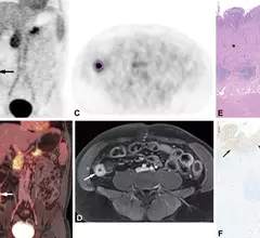
PET/MRI of FAPI radiotracer uptake in Crohn's disease

New imaging technique could change how Crohn's disease is treated

The FAPI radiotracer experts used in this research can bind specifically to the connective tissue cells that cause intestinal wall fibrosis common to Crohn's disease.

Around the web

Positron, a New York-based nuclear imaging company, will now provide Upbeat Cardiology Solutions with advanced PET/CT systems and services. 

The nuclear imaging isotope shortage of molybdenum-99 may be over now that the sidelined reactor is restarting. ASNC's president says PET and new SPECT technologies helped cardiac imaging labs better weather the storm.

CMS has more than doubled the CCTA payment rate from $175 to $357.13. The move, expected to have a significant impact on the utilization of cardiac CT, received immediate praise from imaging specialists.

Trimed Popup
Trimed Popup