Magnetic Resonance Imaging

Magnetic resonance imaging (MRI) is the gold standard imaging modality for soft tissues. It produces detail cross-sectional images of soft tissue and bone anatomy, including muscles, tendons, ligaments, brain and organs, without the use of ionizing radiation. In addition to orthopedic imaging, MRI is also used for heart, brain and breast. MRI uses gadolinium contrast in many exams to highlight tissues and blood vessels, which enhances images and offers better diagnostic quality. It can also be used in conjunction with PET scans. How does MRI work? MR creates images by using powerful magnets to polarize hydrogen atoms in water (the body is made of of more than 80% water) so they face in one direction. A radiofrequency pulse is then used to ping these atoms, causing them to wobble, or resonate. The MRI coils detect this and computers can assemble images from the signals. Basic MRI scans will focus on the resonance of fat and water in two different sequences, which highlight and contrast different features in the anatomy.

Thumbnail

When GBCA use in musculoskeletal imaging is and is not appropriate

Although gadolinium-based contrast agents are largely considered safe and are routinely used for MRI exams, experts suggest that providers should still utilize GBCAs sparingly for musculoskeletal studies.

Thumbnail

Prostate cancer size is frequently underestimated on imaging

Newly published data recently revealed just how common the extent of prostate cancer is underestimated based on imaging measurements. 

University of Minnesota team develops compact portable MRI

Curbside MRI: Experts are developing system small enough to fit in a pickup

Someday, getting an MRI exam could be as simple as having food delivered to your door—at least that is the hope of a group of experts at the University of Minnesota who are working on a compact system said to be small enough to sit in the bed of a truck.

Philips' latest collaboration could make xenon MR imaging more widely available

In alliance with Polarean, Philips is rolling out a new MR system with advanced multi-nuclei imaging capabilities that will offer radiologists a detailed evaluation of lung ventilation using xenon gas as a contrast agent.

Incomplete neurovascular imaging work-ups to blame for subsequent stroke in patients with TIA

Up to 37% of patients presenting with transient ischemic attack do not receive a full neurovascular imaging work-up, according to new data published in the American Journal of Roentgenology. 

Thumbnail

AI tool helps identify 'invisible' head injuries on MRIs of college athletes

The tool uses a machine learning technique to identify changes on brain MRIs that would otherwise be overlooked by radiologists due to the subtlety of alterations. 

osteomyelitis on MRI

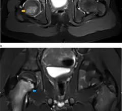
Diagnosing osteomyelitis with abbreviated noncontrast MRI protocols

Using abbreviated MRI protocols for pediatric exams offers similar quality as standard protocols but decreases the need for sedation. 

FDA report details MRI accident caused by sex toy

An FDA adverse event report suggests that although the patient involved in the accident was screened for metal prior to undergoing her MRI, she did not disclose the presence of a “butt plug." 

Around the web

Positron, a New York-based nuclear imaging company, will now provide Upbeat Cardiology Solutions with advanced PET/CT systems and services. 

The nuclear imaging isotope shortage of molybdenum-99 may be over now that the sidelined reactor is restarting. ASNC's president says PET and new SPECT technologies helped cardiac imaging labs better weather the storm.

CMS has more than doubled the CCTA payment rate from $175 to $357.13. The move, expected to have a significant impact on the utilization of cardiac CT, received immediate praise from imaging specialists.

Trimed Popup
Trimed Popup