Magnetic Resonance Imaging

Magnetic resonance imaging (MRI) is the gold standard imaging modality for soft tissues. It produces detail cross-sectional images of soft tissue and bone anatomy, including muscles, tendons, ligaments, brain and organs, without the use of ionizing radiation. In addition to orthopedic imaging, MRI is also used for heart, brain and breast. MRI uses gadolinium contrast in many exams to highlight tissues and blood vessels, which enhances images and offers better diagnostic quality. It can also be used in conjunction with PET scans. How does MRI work? MR creates images by using powerful magnets to polarize hydrogen atoms in water (the body is made of of more than 80% water) so they face in one direction. A radiofrequency pulse is then used to ping these atoms, causing them to wobble, or resonate. The MRI coils detect this and computers can assemble images from the signals. Basic MRI scans will focus on the resonance of fat and water in two different sequences, which highlight and contrast different features in the anatomy.

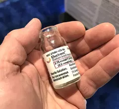
The new MRI contrast agent gadopiclenol, sold under the trade names Elucirem and Vueway by Bracco and Guerbet, used 50% less gadolinium that current MRI agents. #RSNA22 #RSNA

New contrast agent earns positive safety classification from ACR

The ACR Committee on Drugs and Contrast Media stated that exposure to the gadolinium-based contrast agent represents a “sufficiently low or possibly nonexistent” risk to patients.

Thumbnail

Prenatal MRI shines light on the implications of maternal stress during pregnancy

These findings warrant serious consideration by physicians caring for pregnant women in the future, as the link between poor mental health and placental development remains “underappreciated.”

Learning app improves reader performance for prostate MRI

Interactive teaching app improves reader performance on prostate MRI

Experts involved in the app’s creation suggested that because it was designed to mimic real life, its use among emerging radiologists could potentially help address the issue of subjectivity and reader variability in interpreting mpMRI scans of the prostate.

Thumbnail

Radioactive substances unnecessary in new method for measuring brain glucose metabolism

Rather than administering radiolabeled glucose for exams, imagers give patients a small amount of a harmless glucose solution that is said to be equivalent to a can of a carbonated drink.

Thumbnail

MRI and PET findings could guide treatment of lingering concussion symptoms

Abnormalities of the thalamus could be to blame for persistent symptoms in the months following a concussion, according to new research.

COVID-19 coronavirus mask smell

COVID long-haulers with loss of smell display brain connectivity impairments on MRI

Experts recently identified alterations in regions of the brain that process smell in COVID long-haulers. 

Nearly 60% of YouTube videos about MRI claustrophobia are deemed 'useless'

Nonprofessionals are responsible for nearly 75% of uploaded YouTube videos on the topic, the quality and reliability of which some experts find concerning, according to a new paper published in Cureus.

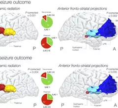
brain connections that cause epileptic seizures

Experts use MRI to identify brain pathways responsible for epileptic seizures

Patients who have these pathways disconnected record marked seizure-free periods of time in the long-term, according to new research.

Around the web

Positron, a New York-based nuclear imaging company, will now provide Upbeat Cardiology Solutions with advanced PET/CT systems and services. 

The nuclear imaging isotope shortage of molybdenum-99 may be over now that the sidelined reactor is restarting. ASNC's president says PET and new SPECT technologies helped cardiac imaging labs better weather the storm.

CMS has more than doubled the CCTA payment rate from $175 to $357.13. The move, expected to have a significant impact on the utilization of cardiac CT, received immediate praise from imaging specialists.

Trimed Popup
Trimed Popup