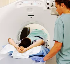
Magnetic Resonance Imaging

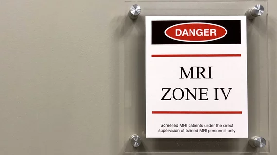
Magnetic resonance imaging (MRI) is the gold standard imaging modality for soft tissues. It produces detail cross-sectional images of soft tissue and bone anatomy, including muscles, tendons, ligaments, brain and organs, without the use of ionizing radiation. In addition to orthopedic imaging, MRI is also used for heart, brain and breast. MRI uses gadolinium contrast in many exams to highlight tissues and blood vessels, which enhances images and offers better diagnostic quality. It can also be used in conjunction with PET scans. How does MRI work? MR creates images by using powerful magnets to polarize hydrogen atoms in water (the body is made of of more than 80% water) so they face in one direction. A radiofrequency pulse is then used to ping these atoms, causing them to wobble, or resonate. The MRI coils detect this and computers can assemble images from the signals. Basic MRI scans will focus on the resonance of fat and water in two different sequences, which highlight and contrast different features in the anatomy.

Nebraska man attempts to escape police custody during MRI

The man was in custody after leading police on a high-speed chase the day before.

Motion-Corrected MRI Scan

MRI results affected by movement? MIT researchers have an AI-powered solution

An MRI's sensitivity to movement during scans often leads to image artifacts, risking misdiagnoses and redundant scans. But a new deep learning model may be able to compensate.

U.S. News & World Report children’s hospitals cardiology heart surgery

Imagers trim pediatric sedation and anesthesia over 11-year period

Continued improvement in imaging techniques and development of clinical practice guidelines may further reduce the need, experts noted. 

Thumbnail

Generic substitute for gadolinium-based contrast agent Gadavist hits the market

The contrast agent, simply called Gadobutrol Injection, has been approved by the U.S. Food and Drug Administration and is now available in the United States.

 background parenchyma enhancement breast MRI

MRI finding linked to heightened cancer risk among women with very dense breasts

A new AI model helped researchers identify a relationship between background parenchyma enhancement on breast MRI exams and breast cancer risk.

artificial intelligence consultation

New-look AI model for imaging results provides its own second opinions

The "dual-view" algorithm is separated into two distinct parts that can learn from one another and catch potential errors. 

Thumbnail

PROMISE trial results reinforce confidence in new MRI contrast agent

The randomized, double-blinded phase 3 study revealed gadopiclenol to be noninferior to gadobutrol for all qualitative visualization parameters as rated by all blinded readers involved in the trial.

Thumbnail

Experts argue for the use of MRI in all first-episode psychosis patients

A new meta-analysis revealed that up to 26% of FEP patients display intracranial radiological abnormalities on MRI, some of which can alter clinical management.

Around the web

Positron, a New York-based nuclear imaging company, will now provide Upbeat Cardiology Solutions with advanced PET/CT systems and services. 

The nuclear imaging isotope shortage of molybdenum-99 may be over now that the sidelined reactor is restarting. ASNC's president says PET and new SPECT technologies helped cardiac imaging labs better weather the storm.

CMS has more than doubled the CCTA payment rate from $175 to $357.13. The move, expected to have a significant impact on the utilization of cardiac CT, received immediate praise from imaging specialists.