Magnetic Resonance Imaging

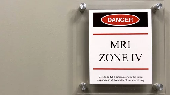
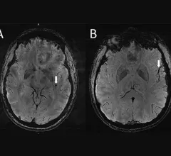
Magnetic resonance imaging (MRI) is the gold standard imaging modality for soft tissues. It produces detail cross-sectional images of soft tissue and bone anatomy, including muscles, tendons, ligaments, brain and organs, without the use of ionizing radiation. In addition to orthopedic imaging, MRI is also used for heart, brain and breast. MRI uses gadolinium contrast in many exams to highlight tissues and blood vessels, which enhances images and offers better diagnostic quality. It can also be used in conjunction with PET scans. How does MRI work? MR creates images by using powerful magnets to polarize hydrogen atoms in water (the body is made of of more than 80% water) so they face in one direction. A radiofrequency pulse is then used to ping these atoms, causing them to wobble, or resonate. The MRI coils detect this and computers can assemble images from the signals. Basic MRI scans will focus on the resonance of fat and water in two different sequences, which highlight and contrast different features in the anatomy.

veterans_counseling.jpg

Veterans who experience chronic pain and trauma have connectivity abnormalities on brain imaging

Though it remains unclear whether these patterns are a result of pain and trauma or if they indicate that a person is more susceptible to symptoms, authors noted that these objective measures could help personalize treatment plans.

May 10, 2022
How radiology should prepare for AUC clinical decision support reporting requirements. CMS to require AUC CDS January 1, 2023.

How radiology should prepare for AUC clinical decision support reporting requirements

An overview for the coming January 2023 mandate to use clinical decision support for all advanced imaging orders.

May 10, 2022
With the shortage, which pertains specifically to all formulations and concentrations of GE Healthcare’s Omnipaque (iohexol), expected to last until mid-June, several organizations have eluded that conservation efforts are of critical importance.#contrastshortage #CTcontrast #iodinatedcontrast

Preserving contrast media supplies: 7 ACR recommendations

With the shortage, which pertains specifically to all formulations and concentrations of GE Healthcare’s Omnipaque (iohexol), expected to last until mid-June, several organizations have alluded that conservation efforts are of critical importance.

May 9, 2022
Functional magnetic resonance imaging can offer providers insight into brain connectivity abnormalities and how they correlate with cognitive impairments observed in patients with insomnia. #insomnia #troublesleeping

Connectivity abnormalities observed on MRIs of insomnia patients

The authors explained that the increased connectivity observed in those with insomnia could lead to hypersensitivity to external stimulation, which might result in a state of hyperarousal. 

May 6, 2022
Safety information for patients taking Aduhelm has been updated by the FDA to include the addition of two MRI scans during the first year of treatment. #alzheimers #alzheimerstreatment

Radiologists should be aware of amyloid-related imaging abnormalities for patients taking Alzheimer's drug

Safety information for patients taking Aduhelm has been updated by the FDA to include the addition of two MRI scans during the first year of treatment.

May 5, 2022
A new artificial intelligence tool can detect heart disease on cardiovascular MRI scans in seconds with equal or superior precision as clinicians. 

Could cardiac MRI become the reference standard for diagnosing heart failure?

Cardiac MRI may offer a more accurate and less invasive method for diagnosing heart failure, a new study published in the European Heart Journal shows. 

May 5, 2022
COVID-19 vaccine associated myocarditis on short-axis 1.5T MRI images of a 19-year-old man who presented with chest pain three days following the second dose of an mRNA COVID-19 vaccine.

New cardiac MRI analysis offers updated insight into long-term impact of vaccine-related myocarditis

Months after their initial myocarditis diagnosis, late gadolinium enhancement (LGE) was observed in 62% of patients on follow-up cardiac MRI.

May 4, 2022

Cardiac implantable electronic devices can be safely reprogrammed to MRI-safe mode via remote operator

In 209 study participants, the remote reprogramming resulted in a successful, event-free MRI scan for every patient.

May 4, 2022

Around the web

Automated AI-generated measurements combined with annotated CT images can improve treatment planning and help referring physicians and patients better understand their disease, explained Sarah Jane Rinehart, MD, director of cardiac imaging with Charleston Area Medical Center.

Two advanced algorithms—one for CAC scores and another for segmenting cardiac chamber volumes—outperformed radiologists when assessing low-dose chest CT scans. 

"Gen AI can help tackle repetitive tasks and provide insights into massive datasets, saving valuable time," Thomas Kurian, CEO of Google Cloud, said Tuesday. 

Trimed Popup
Trimed Popup