Magnetic Resonance Imaging

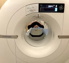
Magnetic resonance imaging (MRI) is the gold standard imaging modality for soft tissues. It produces detail cross-sectional images of soft tissue and bone anatomy, including muscles, tendons, ligaments, brain and organs, without the use of ionizing radiation. In addition to orthopedic imaging, MRI is also used for heart, brain and breast. MRI uses gadolinium contrast in many exams to highlight tissues and blood vessels, which enhances images and offers better diagnostic quality. It can also be used in conjunction with PET scans. How does MRI work? MR creates images by using powerful magnets to polarize hydrogen atoms in water (the body is made of of more than 80% water) so they face in one direction. A radiofrequency pulse is then used to ping these atoms, causing them to wobble, or resonate. The MRI coils detect this and computers can assemble images from the signals. Basic MRI scans will focus on the resonance of fat and water in two different sequences, which highlight and contrast different features in the anatomy.

Thumbnail

MRI scans portray the significant impact of childhood neglect

“Here we show that that the opportunities that a child has in early life to learn and grow will impact not just their behavior but their actual brain development and brain structure for years to come,” authors of the new research shared.

Radiologists are overlooking signs of pancreatic cancer on imaging more and more, new study indicates

The research revealed that 7.7% of patients screened for pancreatic cancer had their tumors missed on initial imaging exams but were diagnosed with cancer between three and 18 months later.  

Thumbnail

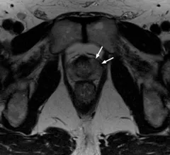
Prostate cancer detection boosted with computer assistance

The addition of computer-aided diagnostic generated MRI series could help radiologists identify clinically significant prostate cancer more frequently. 

Thumbnail

Prostate cancer patients who undergo surveillance MRI in lieu of yearly biopsy not at increased risk

Results of the new work indicate that risks are not exacerbated when imaging is the chosen method of surveillance up until the three-year mark, suggesting that patients can safely forego invasive biopsy for a limited period. 

Thumbnail

Fetal MRI reveals how opioid use during pregnancy limits brain growth in utero

This study is one of the largest to date to assess brain growth in opioid-exposed fetuses in utero. 

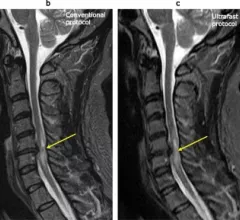
Ultrafast MRI protocol reduces scan time by 10 minutes for cervical imaging

Experts involved in the study suggested that the protocol could open doors leading to greater utilization of MRI in the future. 

Thumbnail

New cardiac MR technique cuts scan time by more than half

Using a new 4D flow cardiac MRI scan, experts were able to complete cardiac examinations in just eight minutes.

New contrast agent receives FDA approval after competitors collaborate on development

The Sept. 21 announcement states that Gadopiclenol Injection—a highly stable macrocyclic gadolinium-based contrast agent—is approved for use in adult and pediatric patients aged 2 years and older during MRI exams.

Around the web

Positron, a New York-based nuclear imaging company, will now provide Upbeat Cardiology Solutions with advanced PET/CT systems and services. 

The nuclear imaging isotope shortage of molybdenum-99 may be over now that the sidelined reactor is restarting. ASNC's president says PET and new SPECT technologies helped cardiac imaging labs better weather the storm.

CMS has more than doubled the CCTA payment rate from $175 to $357.13. The move, expected to have a significant impact on the utilization of cardiac CT, received immediate praise from imaging specialists.

Trimed Popup
Trimed Popup