Magnetic Resonance Imaging

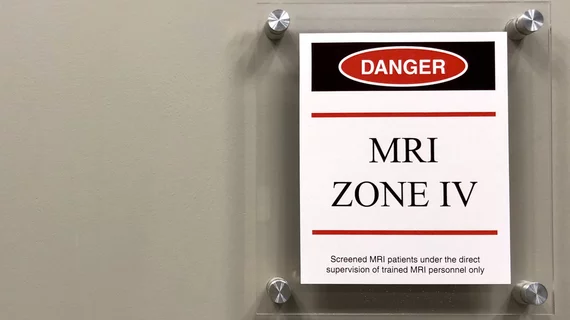
Magnetic resonance imaging (MRI) is the gold standard imaging modality for soft tissues. It produces detail cross-sectional images of soft tissue and bone anatomy, including muscles, tendons, ligaments, brain and organs, without the use of ionizing radiation. In addition to orthopedic imaging, MRI is also used for heart, brain and breast. MRI uses gadolinium contrast in many exams to highlight tissues and blood vessels, which enhances images and offers better diagnostic quality. It can also be used in conjunction with PET scans. How does MRI work? MR creates images by using powerful magnets to polarize hydrogen atoms in water (the body is made of of more than 80% water) so they face in one direction. A radiofrequency pulse is then used to ping these atoms, causing them to wobble, or resonate. The MRI coils detect this and computers can assemble images from the signals. Basic MRI scans will focus on the resonance of fat and water in two different sequences, which highlight and contrast different features in the anatomy.

Thumbnail

Rod Stewart is helping to address 'ridiculous' backlogs by paying for patients' imaging exams

The legendary singer's actions were prompted, in part, by his own experience undergoing medical imaging recently.

0.55T MRI lumbar compared to 1.5T sequence #lumbarMRI #1.5T

Low-field MRI systems improve accessibility, but what kind of diagnostic quality do they offer?

Experts recently compared the use of a 0.55T low-field MRI system to a 1.5T system to determine whether the results rendered were within an acceptable diagnostic range.

Thumbnail

Fatal accident calls MRI safety standards into question—are more regulations needed?

There are a number of well-intended recommendations for preventing MRI injuries, but many of them stop short of implementing safety requirements.

Thumbnail

Some long COVID patients display 'severe' changes in their brain

For patients with anxiety and depression, even mild cases of long COVID could result in functional and structural alterations of the brain.

Thumbnail

Expert left 'astounded' by gadolinium discovery

New data out of the University of New Mexico suggest that the imaging agent’s physiologic reach may exceed what was previously believed.

long covid lung CT

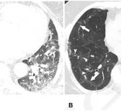
Some long COVID patients continue to display multi-organ damage one year after recovery

A new study utilizing multi-organ MRI scans recently identified organ impairment in 62% of COVID long haulers six months after their initial diagnosis; 29% of these individuals continued to display damage in at least one organ at the 12-month mark.

#footdrop #THAcomplications #THA

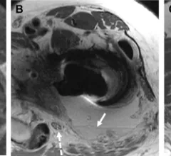
Common MRI findings in 'foot drop' cases

A new paper in the European Journal of Radiology offers an updated overview of common MRI findings in patients who develop foot drop following total hip arthroplasty.

Thumbnail

New contrast agent is officially available for use in hospitals and clinics

A Feb. 14 announcement from Bracco indicates that the recently FDA-approved, gadolinium-based contrast agent VUEWAY, also known as gadopiclenol, is officially being used for patients undergoing MR imaging.

Around the web

Positron, a New York-based nuclear imaging company, will now provide Upbeat Cardiology Solutions with advanced PET/CT systems and services. 

The nuclear imaging isotope shortage of molybdenum-99 may be over now that the sidelined reactor is restarting. ASNC's president says PET and new SPECT technologies helped cardiac imaging labs better weather the storm.

CMS has more than doubled the CCTA payment rate from $175 to $357.13. The move, expected to have a significant impact on the utilization of cardiac CT, received immediate praise from imaging specialists.

Trimed Popup
Trimed Popup