Magnetic Resonance Imaging

Magnetic resonance imaging (MRI) is the gold standard imaging modality for soft tissues. It produces detail cross-sectional images of soft tissue and bone anatomy, including muscles, tendons, ligaments, brain and organs, without the use of ionizing radiation. In addition to orthopedic imaging, MRI is also used for heart, brain and breast. MRI uses gadolinium contrast in many exams to highlight tissues and blood vessels, which enhances images and offers better diagnostic quality. It can also be used in conjunction with PET scans. How does MRI work? MR creates images by using powerful magnets to polarize hydrogen atoms in water (the body is made of of more than 80% water) so they face in one direction. A radiofrequency pulse is then used to ping these atoms, causing them to wobble, or resonate. The MRI coils detect this and computers can assemble images from the signals. Basic MRI scans will focus on the resonance of fat and water in two different sequences, which highlight and contrast different features in the anatomy.

Thumbnail

'Significant' brain abnormalities shown to persist 6 months after COVID recovery

The regions that displayed the most significant differentiations in susceptibility values are known to be associated with neurological conditions such as fatigue, insomnia, anxiety, depression, headaches and other cognitive issues—all common complaints among many COVID long haulers. 

How safe is 7T MRI for patients with neurosurgical implants?

These devices have been tested with lower MRI fields, like 1.5T and 3T, but less is known about their compatibility with more powerful 7T equipment. 

Thumbnail

Experts discover brain connectivity patterns unique to children with autism

The findings were also found to be associated with the severity of symptoms in individuals with ASD.

Thumbnail

New lesion measurement better predicts MS progression

Researchers recently presented a new method that considers how already existing lesions grow over time as an indicator of disease progression.

Thumbnail

New study may give insight into the neurologic dysfunction of some long-COVID patients

Vessel wall imaging—an MRI technique that offers high resolution images of the vessel wall—may hold clues as to the mechanisms by which COVID infections continue to cause neurologic symptoms after initial recovery in some patients.  

A convicted murderer's fate could hinge on different interpretations of her brain scans

In the case of a convicted murderer, a New York neuro specialist cited a slew of neuroimaging findings that indicate “severe dysfunction” that could have numbed the defendant’s “brakes of inhibition.” 

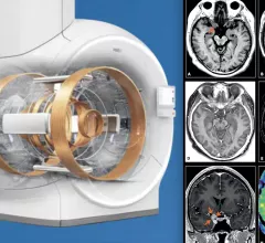
MRI system advances include helium-free MRI systems (left is Philips version of this system), and compressed sensing (right, GE's version of this technology), which can greatly reduce scan times. Both technologies were mentioned in an overview by Signet Research of new tech that will be at RSNA 2022.

VIDEO: Overview of MRI market and technology trends

Bhvita Jani, research manager, Signify Research, explains some key trends and technology advances in the magnetic resonance imaging (MRI) market leading into the Radiological Society of North America (RSNA) 2022 meeting. 

Thumbnail

Top MRI trends include helium-free systems, compressed sensing and AI

Signify Research outlines several key technologies and trends in MRI to look for at the Radiological Society of North America 2022 meeting.

Around the web

Positron, a New York-based nuclear imaging company, will now provide Upbeat Cardiology Solutions with advanced PET/CT systems and services. 

The nuclear imaging isotope shortage of molybdenum-99 may be over now that the sidelined reactor is restarting. ASNC's president says PET and new SPECT technologies helped cardiac imaging labs better weather the storm.

CMS has more than doubled the CCTA payment rate from $175 to $357.13. The move, expected to have a significant impact on the utilization of cardiac CT, received immediate praise from imaging specialists.

Trimed Popup
Trimed Popup