Magnetic Resonance Imaging

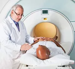
Magnetic resonance imaging (MRI) is the gold standard imaging modality for soft tissues. It produces detail cross-sectional images of soft tissue and bone anatomy, including muscles, tendons, ligaments, brain and organs, without the use of ionizing radiation. In addition to orthopedic imaging, MRI is also used for heart, brain and breast. MRI uses gadolinium contrast in many exams to highlight tissues and blood vessels, which enhances images and offers better diagnostic quality. It can also be used in conjunction with PET scans. How does MRI work? MR creates images by using powerful magnets to polarize hydrogen atoms in water (the body is made of of more than 80% water) so they face in one direction. A radiofrequency pulse is then used to ping these atoms, causing them to wobble, or resonate. The MRI coils detect this and computers can assemble images from the signals. Basic MRI scans will focus on the resonance of fat and water in two different sequences, which highlight and contrast different features in the anatomy.

Thumbnail

How maternal COVID-related stress can alter fetal brain development

The preliminary results of a new study published in the medRxiv preprint server highlight variances in fetal brain development based on mothers’ perceived stress during the onset of COVID.

MRI shows structural, functional brain abnormalities in Lyme disease long-haulers

Compared to a group of patients who had not been previously diagnosed with Lyme disease, those who had been infected displayed unusual activity in the frontal lobe of the brain on functional MRI scans.

Structural brain abnormalities linked to functional seizures

Using MRI imaging, the experts found that patients who experienced functional seizures had thinning in the superior temporal cortex and thickening of the left occipital cortex.

FDA greenlights AI-powered MR software that could give radiotherapy planning a boost

The U.S. Food and Drug Administration granted Philips 510(k) clearance for its AI-powered MRI platform tailored to the treatment of head and neck cancers. 

Helium shortage prompts new sense of urgency in the medical imaging community

In recent years the world’s supply of helium has raised heightened concerns, and some suppliers recently started to ration the nonrenewable element. 

Triplet pregnancy fetus in fetu: A rare case report

While FIF is an extremely rare condition—occurring in about one in 500,000 births—it is even more rare in triplet pregnancies.

Brain scans reveal single doses of nicotine inhibiting estrogen production in women

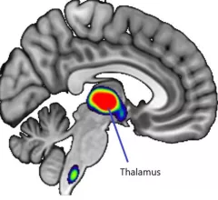
On MRI and PET brain scans, women who were exposed to a small dose of nicotine showed altered activity in the thalamus region of the brain.

Thumbnail

What is the radiologist's role in variations of prostate cancer detection?

Prior studies have focused on radiologist performance rather than patient outcomes, leaving the topic of variable diagnoses and what factors impact them—race, ethnicity, age, biopsy type, etc.—open for debate. 

Around the web

Positron, a New York-based nuclear imaging company, will now provide Upbeat Cardiology Solutions with advanced PET/CT systems and services. 

The nuclear imaging isotope shortage of molybdenum-99 may be over now that the sidelined reactor is restarting. ASNC's president says PET and new SPECT technologies helped cardiac imaging labs better weather the storm.

CMS has more than doubled the CCTA payment rate from $175 to $357.13. The move, expected to have a significant impact on the utilization of cardiac CT, received immediate praise from imaging specialists.

Trimed Popup
Trimed Popup