Magnetic Resonance Imaging

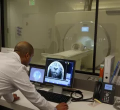
Magnetic resonance imaging (MRI) is the gold standard imaging modality for soft tissues. It produces detail cross-sectional images of soft tissue and bone anatomy, including muscles, tendons, ligaments, brain and organs, without the use of ionizing radiation. In addition to orthopedic imaging, MRI is also used for heart, brain and breast. MRI uses gadolinium contrast in many exams to highlight tissues and blood vessels, which enhances images and offers better diagnostic quality. It can also be used in conjunction with PET scans. How does MRI work? MR creates images by using powerful magnets to polarize hydrogen atoms in water (the body is made of of more than 80% water) so they face in one direction. A radiofrequency pulse is then used to ping these atoms, causing them to wobble, or resonate. The MRI coils detect this and computers can assemble images from the signals. Basic MRI scans will focus on the resonance of fat and water in two different sequences, which highlight and contrast different features in the anatomy.

Thumbnail

Neuroimaging analysis suggests that the brains of patients with depression are not so different after all

“Results of this case-control study suggest that even for maximum univariate biological differences, deviations between patients with MDD and healthy controls were remarkably small," experts shared in JAMA Psychiatry.

soft tissue lymphoma versus soft tissue tumor

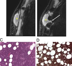
Soft tissue lymphomas versus soft tissue tumors: MRI features reliably differentiate between the two

A new analysis offers a detailed comparison of soft-tissue lymphomas and soft-tissue tumors based on imaging characteristics from MRI scans—an area of study that has not yet been rigorously explored, the authors of the paper indicated.

bone and soft tissue tumors

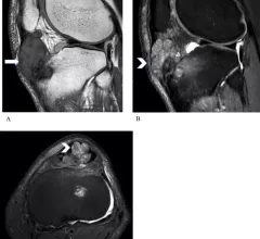
Radiologists' understanding of tumor 'mimics' vital when assessing bone and soft tissue lesions

Authors of the new EJR paper explained that, although most of these tumors present in a similar way—with a lump or localized pain—their origins are wide-ranging and require the use of additional imaging to characterize the lesion. 

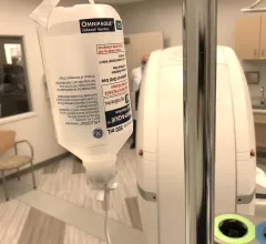
Omnipaque and Visipaque still listed among the FDA's drug shortages

According to the FDA’s most recent update, some presentations will remain out of stock or backordered until January 2023. 

Thumbnail

Time-of-flight intracranial MRA at 5T comparable to 7T, new analysis shows

A new paper published in Radiology details an analysis of 3-T, 5T and 7T MRI to determine which system could achieve the best intracranial vascular image quality.

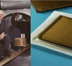
Two of the top stories this month included the death of MRI pioneer Raymond Vahan Damadian. Experts at MIT also developed ultrasound stickers that can be worn in the same manner as a Band-Aid while also producing diagnostic quality images in real-time. #MRI

Top Health Imaging stories in August 2022

Here are the most popular stories on Health Imaging in August, based on our website analytics. 

MRI exams reveal the impact of steroid use on the brain

Experts involved in the study suggest that their findings could help explain the neuropsychiatric effects seen in many patients who are prescribed steroids long-term.

WTC first responders

Can PTSD cause dementia? Imaging of 9/11 first responders suggests it's possible

Compared to World Trade Center responders who have not been diagnosed with PTSD, those who had received the diagnosis were observed to have decreased fractional anisotropy of white matter tracts on their brain MRIs.

Around the web

Positron, a New York-based nuclear imaging company, will now provide Upbeat Cardiology Solutions with advanced PET/CT systems and services. 

The nuclear imaging isotope shortage of molybdenum-99 may be over now that the sidelined reactor is restarting. ASNC's president says PET and new SPECT technologies helped cardiac imaging labs better weather the storm.

CMS has more than doubled the CCTA payment rate from $175 to $357.13. The move, expected to have a significant impact on the utilization of cardiac CT, received immediate praise from imaging specialists.