Magnetic Resonance Imaging

Magnetic resonance imaging (MRI) is the gold standard imaging modality for soft tissues. It produces detail cross-sectional images of soft tissue and bone anatomy, including muscles, tendons, ligaments, brain and organs, without the use of ionizing radiation. In addition to orthopedic imaging, MRI is also used for heart, brain and breast. MRI uses gadolinium contrast in many exams to highlight tissues and blood vessels, which enhances images and offers better diagnostic quality. It can also be used in conjunction with PET scans. How does MRI work? MR creates images by using powerful magnets to polarize hydrogen atoms in water (the body is made of of more than 80% water) so they face in one direction. A radiofrequency pulse is then used to ping these atoms, causing them to wobble, or resonate. The MRI coils detect this and computers can assemble images from the signals. Basic MRI scans will focus on the resonance of fat and water in two different sequences, which highlight and contrast different features in the anatomy.

how_secure_is_that_scanner.jpg

How Secure Is That Scanner?

In a world of networked medical devices, it’s not hard to imagine a radiology-heavy cyberattack that is not only malicious but also ingenious.
 

June 25, 2019

SBRT an effective local treatment for HCC patients

“The results of this research are extremely exciting, as it will significantly impact clinical care,” reported study author Mishal Mendiratta-Lala, MD, with the division of abdominal radiology at Michigan Medicine in Ann Arbor.

May 10, 2019
""

Cardiac MRI-derived T2 mapping may help heart failure patients

Researchers used T2 mapping taken from weekly cardiac MRIs to help identify cardiotoxicity at an early stage, according to results of a pig study published Feb. 18 in the Journal of the American College of Cardiology. The findings could help cancer patients at risk of chemo-induced heart failure.

February 22, 2019

T2 mapping may uncover cardiotoxic marker early enough to prevent heart failure

T2 mapping derived from weekly cardiac MRIs helped researchers identify cardiotoxicity at an early and reversible stage, a finding which may have implications for cancer patients at risk of chemotherapy-induced heart failure.

February 21, 2019
Money

Insurer's price calculator severely underestimates breast MRI cost for Philadelphia woman

Although many insurance providers have invested in price calculators for patients to determine out-of-pocket costs, one Philadelphia woman discovered they may severely underestimate actual prices for services, according to a recent report by The Philadelphia Inquirer.

January 10, 2019

Philips unveils new radiation oncology portfolio for a more confident path to treatment at ASTRO 2018

New Big Bore RT and Ingenia Ambition/Elition MR-RT imaging systems and Pinnacle Evolution treatment planning software support improved patient care, accelerated time to treatment and enhanced patient satisfaction.

October 23, 2018

Can diffusion-weighted imaging supplement dynamic contrast-enhanced breast MRI?

Diffusion-weighted imaging (DWI) could help differentiate between benign and malignant tumors and predict tumor recurrence in breast cancer patients, according to research published Oct. 4 in Academic Radiology. The authors noted that DWI may also serve as a beneficial supplement to contrast-enhanced breast MRI.

October 5, 2018
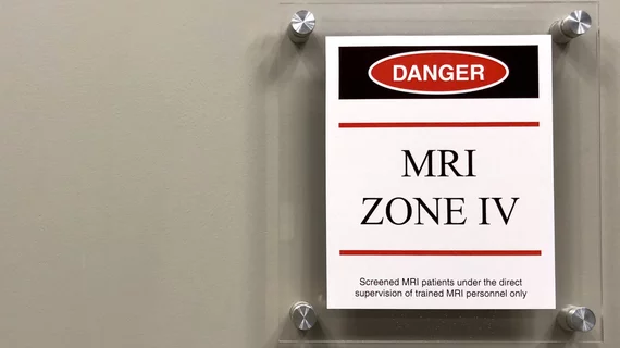
A Long Island, New York, man was arrested last month after a handgun he possessed fired and injured him while in an MRI room, according to a June 20 report from the LI Herald.

New York patient discharges gun in MRI room, charged with reckless endangerment

A Long Island, New York, man was arrested last month after a handgun he possessed fired and injured him while in an MRI room, according to a June 20 report from the LI Herald.

June 21, 2018

Around the web

Automated AI-generated measurements combined with annotated CT images can improve treatment planning and help referring physicians and patients better understand their disease, explained Sarah Jane Rinehart, MD, director of cardiac imaging with Charleston Area Medical Center.

Two advanced algorithms—one for CAC scores and another for segmenting cardiac chamber volumes—outperformed radiologists when assessing low-dose chest CT scans. 

"Gen AI can help tackle repetitive tasks and provide insights into massive datasets, saving valuable time," Thomas Kurian, CEO of Google Cloud, said Tuesday. 

Trimed Popup
Trimed Popup